. 2005 Jun 1;80(5):727-37.
doi: 10.1002/jnr.20501.

Pontine-wave generator activation-dependent memory processing of avoidance learning involves the dorsal hippocampus in the rat

Affiliations

Pontine-wave generator activation-dependent memory processing of avoidance learning involves the dorsal hippocampus in the rat

Subimal Datta et al. J Neurosci Res. .

Abstract

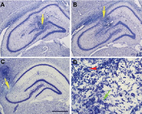
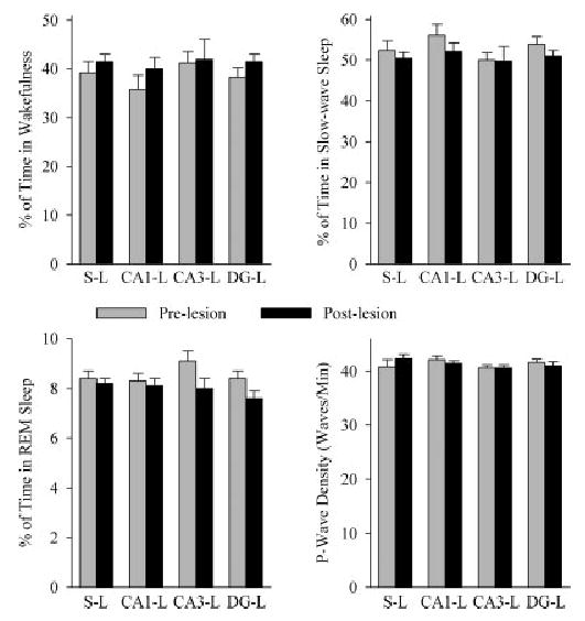
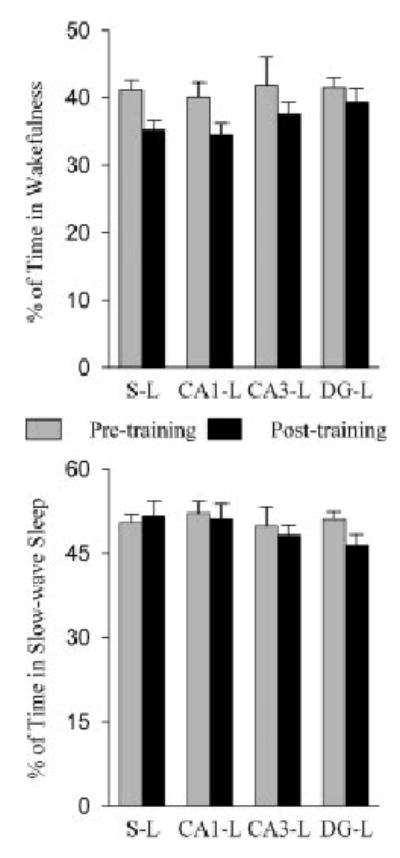
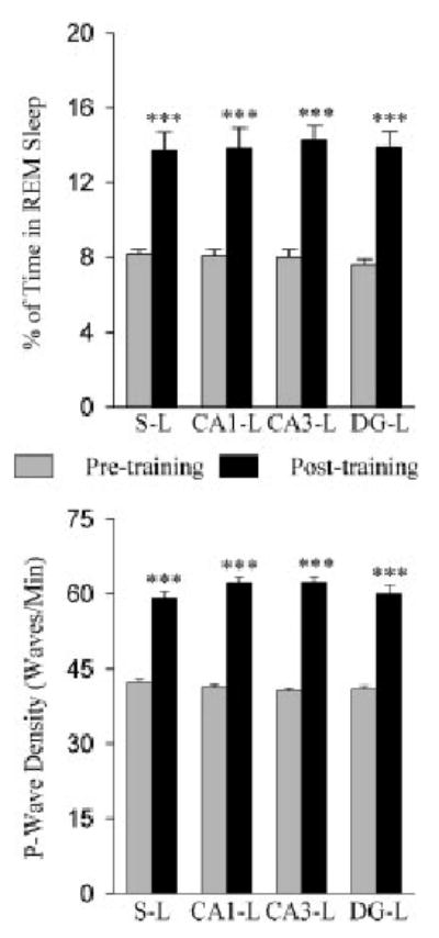
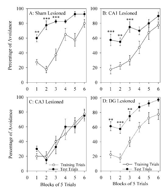
The aim of this study was to test the hypothesis that the dorsal hippocampus plays a critical role in pontine-wave (P-wave) generator activation-dependent memory processing of two-way active avoidance (TWAA) learning. To achieve this objective, rats were given small bilateral lesions in the CA1, dentate gyrus (DG), or CA3 region of the dorsal hippocampus by microinjecting ibotenic acid. After recovery, lesioned and sham-lesioned rats were trained on a TWAA learning paradigm, allowed a 6-hr period of undisturbed sleep, and then were tested on the same TWAA paradigm. It was found that lesions in the CA3 region impaired retention of avoidance learning. Conversely, lesions in the CA1 and DG regions had no effect on TWAA learning retention. None of the groups showed any changes in the baseline sleep-wake cycle or in the acquisition of TWAA learning. All rats showed increased rapid eye movement (REM) sleep and increased REM sleep P-wave density during the subsequent 6-hr recording period. Impaired retention in the CA3 group occurred despite an increase in REM sleep and P-wave density, suggesting that during REM sleep, the P-wave generator interacts with the CA3 region of the dorsal hippocampus to aid in consolidation of TWAA learning. The results of the present study thus demonstrate that P-wave generator activation-dependent consolidation of memory requires an intact CA3 subfield of the dorsal hippocampus. The results also provide evidence that under mnemonic pressure, the dorsal hippocampus may not be involved directly in regulating the sleep-wake cycle.


Figures

Fig. 1
Fig. 1
Examples of histologic localization of ibotenic acid microin-jection-induced lesion sites in different subfields of the dorsal hippocampus. A: A representative coronal section from a rat brain showing an ibotenic acid microinjection-induced lesion in the dentate gyrus (yellow arrow). B: A representative coronal section showing an ibotenic acid microinjection-induced lesion in the CA1 (yellow arrow). C: A representative coronal section showing an ibotenic acid micro-injection-induced lesion in the CA3 (yellow arrow). D: A magnified photomicrograph of part of the ibotenic acid diffusion site in the CA3 subfield demonstrating the cell loss caused by ibotenic acid microinjection. The red arrowhead points to the core of the ibotenic acid diffusion site showing the invasion of glial cells caused by the degeneration of neuronal cells. Note the presence of normal intact cell bodies (green arrow in D) about 400 μm away from the center of the lesion site. Scale bar = 1 mm (A–C); 200 μm (D).
Fig. 2
Fig. 2
The effects of lesioning different subfields of the dorsal hippocampus on sleep–wake parameters. Bars represent the total percentage (mean and SEM) of time spent in wakefulness, slow-wave sleep, or rapid eye movement (REM) sleep, or the pontine-wave (P-wave) density during REM sleep in the 6-hr sleep–wake recording sessions before (light gray bars) and after (dark gray bars) lesioning. Note that ibotenic acid microinjection-induced lesions and sham-lesions did not change the total percentage of wakefulness, slow-wave sleep, or REM sleep, nor did it change REM sleep P-wave density. S-L, sham lesioned group; CA1-L, CA1 lesioned group; CA3-L, CA3 lesioned group; DG-L, dentate gyrus lesioned group.
Fig. 3
Fig. 3
Effects of two-way active avoidance (TWAA) training trials on wakefulness and slow-wave sleep in rats with sham-lesions and lesions in the different subfields of the dorsal hippocampus. Bars represent the total percentage (mean and SEM) of time in wakefulness or slow-wave sleep during the 6-hr sleep recording sessions. Note that the percentages of wakefulness and slow-wave sleep during the 6-hr sleep–wake recording session after training trials are comparable to the percentages of wakefulness and slow-wave sleep during the baseline recording session. Abbreviations are the same as in Figure 2.
Fig. 4
Fig. 4
Effects of two-way active avoidance (TWAA) training trials on rapid eye movement (REM) sleep and REM sleep pontine-wave (P-wave) density in animals with sham-lesions and lesions in the different subfields of the dorsal hippocampus. In the upper panel, bars represent the total percentage (mean and SEM) of time spent in REM sleep during the 6-hr sleep recording session. In the lower panel, bars represent the REM sleep P-wave density (mean and SEM) during the 6-hr recording session. Note that the percentage of REM sleep and the percentage of P-wave density are significantly higher in the recording session after learning training trials than they were during the baseline recording session. Post-hoc tests (Scheffe’s F-test); ***P < 0.001 for comparison between baseline and post-training recording sessions.
Fig. 5
Fig. 5
Learning curves of the dorsal hippocampus sham-lesioned and lesioned rats. Learning curves for the two-way active avoidance (TWAA) task before (training trials session; empty circle) and after (test trials session; filled circle) 6 hr of undisturbed sleep in the sham-lesioned (A), CA1 lesioned (B), CA3 lesioned (C), and DG lesioned (D) rats. The percentage of successful avoidance (mean and SEM) is plotted for every consecutive six blocks of five trials. Note that the percentages of avoidance in the training trials are comparable between sham, CA1, CA3, and DG lesioned rats. In the first three blocks of test trails, sham-lesioned, CA1 lesioned, and DG lesioned rats’ percentages of avoidance were significantly higher than was the percentage of avoidance in the first three blocks of training trials. In the CA3 lesioned rats, however, the percentages of avoidance during the test trials were not significantly different from those during the training trials. Post-hoc tests (Scheffe’s F-test); **P < 0.01, ***P < 0.001 for the comparison between training and test trials.

References

    1. Alreja M, Wu M, Liu W, Atkins JB, Leranth C, Shanabrough M. Muscarinic tone sustains impulse flow in the septohippocampal GABA but not cholinergic pathway: implications for learning and memory. J Neurosci. 2000;20:8103–8110. - PMC - PubMed
    1. Aznar S, Rasmussen T, Zimmer J. Impaired learning correlates with size of excitotoxic hippocampal CA3 lesions in adult rats, but shows no amelioration by CA3 transplants. Restor Neurol Neurosci. 1998;13:141–151. - PubMed
    1. Bailey EL, Overstreet DH, Crocker AD. Effects of intrahippocam-pal injections of the cholinergic neurotoxin AF64A on open-field activity and avoidance learning in the rat. Behav Neural Biol. 1986;45:263–274. - PubMed
    1. Bannerman DM, Grubb M, Deacon RM, Yee BK, Feldon J, Rawlins JN. Ventral hippocampal lesions affect anxiety but not spatial learning. Behav Brain Res. 2003;139:197–213. - PubMed
    1. Bannerman DM, Yee BK, Good MA, Heupel MJ, Iversen SD, Rawlins JN. Double dissociation of function within the hippocampus: a comparison of dorsal, ventral, and complete hippocampal cytotoxic lesions. Behav Neurosci. 1999;113:1170–1188. - PubMed

Publication types

Substances