Comparative Study
. 2005 Sep 15;390(Pt 3):681-8.
doi: 10.1042/BJ20050548.

Binding and repression of translation of the cognate mRNA by Trichinella spiralis thymidylate synthase differ from the corresponding interactions of the human enzyme

Affiliations
Comparative Study

Binding and repression of translation of the cognate mRNA by Trichinella spiralis thymidylate synthase differ from the corresponding interactions of the human enzyme

Joanna Cieśla et al. Biochem J. .

Retraction in

Abstract

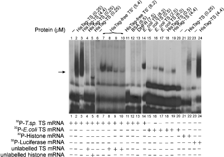
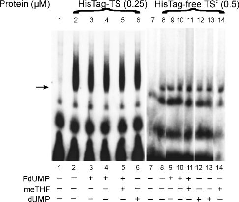
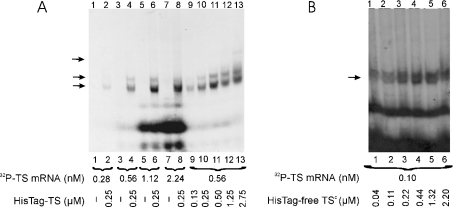
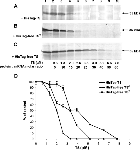
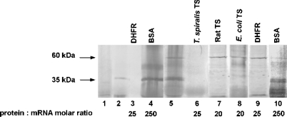
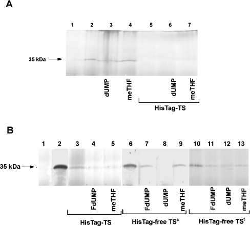
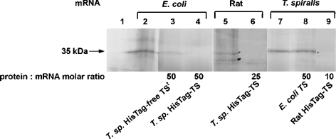
Thymidylate synthase (TS) of Trichinella spiralis, a parasitic nematode causing trichinellosis, was found to bind its own mRNA and repress translation of the latter, similar to its human counter-part [Chu, Koeller, Casey, Drake, Chabner, Elwood, Zinn and Allegra (1991) Proc. Natl. Acad. Sci. U.S.A. 88, 8977-8981]. However, in striking contrast with human TS, the parasite enzyme's interaction with mRNA was not affected by any of the substrate (deoxyuridylate or N(5,10)-methylenetetrahydrofolate) nor by the inhibitor (fluorodeoxyuridylate; used alone or in the presence of N(5,10)-methylenetetrahydrofolate) similar to that shown for the bifunctional enzyme from Plasmodium falciparum [Zhang and Rathod (2002) Science 296, 545-547]. Moreover, repression of the translation of the parasite enzyme was enhanced by the same ligands that were shown by others (Chu et al., 1991) to prevent human TS from impairing its translation. On comparing the capacity of TS to bind to its cognate mRNA, relative to its ability to inhibit its translation, the same enzyme preparation was active as translational repressor at a considerably lower protein/mRNA ratio, suggesting the two phenomena to be disconnected. Of interest is the fact that the presence of the enzyme protein N-terminal methionine proved to be critical for binding, but not for repression of its translation, indicating that mRNA binding requires a methionine or an adduct (i.e. methionine-histidine) at the N-terminus of TS, but that the translational repression effect does not. Notably, chicken liver dihydrofolate reductase, which is incapable of binding to T. spiralis TS mRNA, repressed the translation of TS.


Figures

Figure 1
Figure 1. Binding of T. spiralis and E. coli TS proteins to their own and other mRNAs monitored by the electrophoretic mobility-shift assay
32P-labelled T. spiralis TS mRNA (0.66 nM, 200000 c.p.m. in lanes 1–5 and 11–14 or 0.3 nM, 100 000 c.p.m. in lanes 6–10) was incubated alone (lanes 1 and 6), in the presence of His–TS (HisTag–TS; 0.25 μM in lanes 2, 4 and 5 or 4.4 μM in lane 3), His-tag-free TSc (HisTag-free TSc; complete TS; 5.4 μM; lanes 7–10), His-tag-free TSt (HisTag-free TSt; truncated TS lacking the N-terminal methionine; 8.2 μM; lane 11), BSA (6.5 μM; lane 12), DHFR (7.5 μM; lane 13) or E. coli TS (8.2 μM; lane 14). In the competition experiments, 0.25 μM His–TS was preincubated either with 100 nM unlabelled T. spiralis TS mRNA (lane 4) or unlabelled 100 nM histone mRNA (lane 5), and 5.4 μM His-tag-free TSc was preincubated with 3.2 nM (lane 8), 9.6 nM (lane 9) or 14.4 nM (lane 10) unlabelled T. spiralis TS mRNA before incubation with 32P-labelled T. spiralis TS mRNA. 32P-labelled E. coli TS mRNA (0.25 nM, 300000 c.p.m.; lanes 15–20) was incubated in the absence of protein (lane 20) or in the presence of 0.5 μM (lane 15), 2.5 μM (lane 16) or 6.0 μM (lane 17) E. coli TS, and 0.5 μM (lane 18) or 4.4 μM (lane 19) T. spiralis His–TS. 32P-labelled histone mRNA (0.7 nM, 200000 c.p.m.) was incubated alone (lane 21) or in the presence of 0.25 μM T. spiralis His–TS (lane 22). 32P-labelled luciferase mRNA (0.34 nM, 156000 c.p.m.) was incubated alone (lane 23) or in the presence of 4.4 μM T. spiralis His–TS (lane 24). Position of the RNAse T1-resistant complex is indicated by the arrow.
Figure 2
Figure 2. The effects of dUMP, FdUMP and meTHF on RNA binding activity of T. spiralis TS protein
32P-labelled T. spiralis TS mRNA (0.66 nM, 200000 c.p.m. in lanes 1–6 or 0.64 nM, 100000 c.p.m. in lanes 7–14) was incubated in the absence of protein (lanes 1 and 7) or in the presence of either 0.25 μM His–TS (HisTag–TS; lanes 2–6) or 0.5 μM His-tag-free TSc (HisTag-free TSc; lanes 8–14) without additions (lanes 2 and 8) or together with 25 μM FdUMP (lanes 3 and 9), 250 μM FdUMP (lanes 4 and 10), both 250 μM FdUMP and 250 μM meTHF (lane 5), both 250 μM FdUMP and 500 μM meTHF (lane 11), 25 μM (lane 12) or 250 μM (lanes 6 and 13) dUMP, and 500 μM meTHF (lane 14).
Figure 3
Figure 3. The effect of mRNA and protein concentrations on T. spiralis His–TS protein-TS mRNA (A), and of protein concentration on T. spiralis His-tag-free TSc–TS mRNA complex formation (B), monitored by the electrophoretic mobility-shift assay
32P-labelled TS mRNA was incubated in the absence of protein (A, lanes 1, 3, 5 and 7) or in the presence of 0.25 μM His–TS (HisTag–TS; A, lanes 2, 4, 6 and 8). The mRNA concentrations were as follows: 0.28 nM (A, lanes 1 and 2), 0.56 nM (A, lanes 3 and 4), 1.12 nM (A, lanes 5 and 6) and 2.24 nM (A, lanes 7 and 8). 32P-labelled TS mRNA (0.56 nM, 95000 c.p.m. in A, lanes 9–13 or 0.1 nM, 70000 c.p.m. in B, lanes 1–6) was incubated alone (A, lane 9) or in the presence of increasing concentrations of His–TS (0.13 μM in A, lane 9; 0.25 μM in A, lane 10; 0.50 μM in A, lane 11; 1.25 μM in A, lane 12 and 2.75 μM in A, lane 13) or His-tag-free TSc (HisTag-free TSc; 0.04 μM in B, lane 1; 0.11 μM in B, lane 2; 0.22 μM in B, lane 3; 0.44 μM in B, lane 4; 1.32 μM in B, lane 5 and 2.20 μM in B, lane 6). Positions of the RNAse T1-resistant complex are indicated by the arrows; note a multiband pattern with His–TS.
Figure 4
Figure 4. Dose-dependent inhibition by exogenous T. spiralis His–TS (A), His-tag-free TSc (B) or His-tag-free TSt (C) of T. spiralis TS mRNA translation
(AC) Rabbit reticulocyte lysate was incubated with 6.6 pmol (0.13 μM) T. spiralis TS mRNA (lanes 1–10) and the corresponding TS preparations at concentrations of 0.6 μM (lane 2), 1.3 μM (lane 3), 2.0 μM (lane 4), 2.6 μM (lane 5) and 3.3 μM (lane 6), 3.9 μM (lane 7), 5.2 μM (lane 8), 6.6 μM (lane 9) and 7.8 μM (lane 10). (D) Results of autoradiography assessed by densitometry, expressed in arbitrary units and presented as means±% difference (bars) between the mean and each of the two results. HisTag-TS, His–TS; HisTag-free TS, His-tag-free TS.
Figure 5
Figure 5. The effect of exogenous DHFR, BSA, T. spiralis His–TS, rat His–TS and E. coli TS on in vitro translation of T. spiralis TS and Coleoptera luciferase mRNA
Translation reaction mixtures contained no exogenous mRNA (lane 1), 6.6 pmol (0.13 μM) T. spiralis mRNA (lanes 2–10) and 3.4 pmol (34 nM) luciferase mRNA (lanes 5–10). Exogenously added proteins were 2.6 μM DHFR (lanes 3 and 9), 33 μM BSA (lanes 4 and 10), 3.3 μM T. spiralis His–TS (lane 6), 2.6 μM rat His–TS (lane 7) and 1.3 μM E. coli TS (lane 8).
Figure 6
Figure 6. Influences of dUMP, meTHF and FdUMP on TS protein-inhibited in vitro translation of T. spiralis TS mRNA
(A) Inability of substrates to release enzyme protein-inhibited translation. Each substrate, dUMP (2.5 mM; lanes 3 and 6) or meTHF (600 μM; lanes 4 and 7), was added to the Rabbit Reticulocyte Lysate System containing 6.6 pmol (0.13 μM) T. spiralis TS mRNA (lanes 2–7) with 3.3 μM T. spiralis His–TS (HisTag–TS; lanes 5–7) or without it (lanes 2–4). (B) Enhancement of partial inhibition of translation by His–TS, His-tag-free (HisTag-free) TSc or His-tag-free TSt in the presence of FdUMP or meTHF. FdUMP (50 μM; lanes 4, 7 and 11), dUMP (2.5 mM; lanes 8 and 12) and meTHF (300 μM or 600 μM; lanes 5, 9 and 13 respectively) were added to reaction mixtures containing 6.6 pmol (0.13 μM) T. spiralis TS mRNA (lanes 2–13) and 3.3 μM T. spiralis His–TS (lanes 3–5), His-tag-free TSc (lanes 6–9) or His-tag-free TSt (lanes 10–13).
Figure 7
Figure 7. Effect of various TSs on in vitro translation of mRNA coding the enzyme forms of different specific origin
Translation reactions were incubated without mRNA (lane 1) or with 5.2 pmol (0.1 μM) E. coli TS mRNA (lanes 2–4), and 6.6 pmol (0.13 μM) rat TS mRNA (lanes 5 and 6) or T. spiralis (T. sp.) mRNA (lanes 7–9). Various concentrations of exogenous TSs were added: 5.2 μM T. spiralis His-tag-free TS (HisTag-free TS; lane 3), 5.2 μM (lane 4) or 3.3 μM (lane 6) T. spiralis His–TS (HisTag–TS), 6.6 μM E. coli TS (lane 8) and 1.3 μM rat His–TS (lane 9).

Similar articles

Cited by

References

    1. Carreras C. W., Santi D. The catalytic mechanism and structure of thymidylate synthase. Annu. Rev. Biochem. 1995;64:721–762. - PubMed
    1. Cohen S. S., Flaks J. G., Barner H. D., Loeb M. R., Lichtenstein J. The mode of action of 5-fluorouracil and its derivatives. Proc. Natl. Acad. Sci. U.S.A. 1958;44:1004–1012. - PMC - PubMed
    1. Danenberg P. V. Thymidylate synthetase – a target enzyme in cancer chemotherapy. Biochim. Biophys. Acta. 1977;473:73–97. - PubMed
    1. Hardy L. W., Finer-Moore J. S., Montfort W. R., Jones M. O., Santi D. V., Stroud R. M. Atomic structure of thymidylate synthase: target for rational drug design. Science. 1987;235:448–455. - PubMed
    1. Shoichet B. K., Stroud R. M., Santi D. V., Kuntz I. D., Perry K. M. Structure-based discovery of inhibitors of thymidylate synthase. Science. 1993;259:1445–1450. - PubMed

Publication types

MeSH terms