Electrical impedance along connective tissue planes associated with acupuncture meridians
- PMID: 15882468
- PMCID: PMC1142259
- DOI: 10.1186/1472-6882-5-10
Electrical impedance along connective tissue planes associated with acupuncture meridians
Abstract
Background: Acupuncture points and meridians are commonly believed to possess unique electrical properties. The experimental support for this claim is limited given the technical and methodological shortcomings of prior studies. Recent studies indicate a correspondence between acupuncture meridians and connective tissue planes. We hypothesized that segments of acupuncture meridians that are associated with loose connective tissue planes (between muscles or between muscle and bone) visible by ultrasound have greater electrical conductance (less electrical impedance) than non-meridian, parallel control segments.
Methods: We used a four-electrode method to measure the electrical impedance along segments of the Pericardium and Spleen meridians and corresponding parallel control segments in 23 human subjects. Meridian segments were determined by palpation and proportional measurements. Connective tissue planes underlying those segments were imaged with an ultrasound scanner. Along each meridian segment, four gold-plated needles were inserted along a straight line and used as electrodes. A parallel series of four control needles were placed 0.8 cm medial to the meridian needles. For each set of four needles, a 3.3 kHz alternating (AC) constant amplitude current was introduced at three different amplitudes (20, 40, and 80 microAmps) to the outer two needles, while the voltage was measured between the inner two needles. Tissue impedance between the two inner needles was calculated based on Ohm's law (ratio of voltage to current intensity).
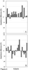
Results: At the Pericardium location, mean tissue impedance was significantly lower at meridian segments (70.4 +/- 5.7 Omega) compared with control segments (75.0 +/- 5.9 Omega) (p = 0.0003). At the Spleen location, mean impedance for meridian (67.8 +/- 6.8 Omega) and control segments (68.5 +/- 7.5 Omega) were not significantly different (p = 0.70).
Conclusion: Tissue impedance was on average lower along the Pericardium meridian, but not along the Spleen meridian, compared with their respective controls. Ultrasound imaging of meridian and control segments suggested that contact of the needle with connective tissue may explain the decrease in electrical impedance noted at the Pericardium meridian. Further studies are needed to determine whether tissue impedance is lower in (1) connective tissue in general compared with muscle and (2) meridian-associated vs. non meridian-associated connective tissue.
Figures


References
-
- Chen KG. Electrical properties of meridians. IEEE Eng Med Biol Mag. 1996;15:58–63. doi: 10.1109/51.499759. - DOI
-
- Hyvarinen J, Karlsson M. Low-resistance skin points that may coincide with acupuncture loci. Med Biol. 1977;55:88–94. - PubMed
-
- Nakatani Y, Yamashita K. Ryodoraku Acupuncture. Tokyo, Ryodoraku Research Institute; 1977.
-
- Nakatani Y. Skin electric resistance and ryodoraku. J Autonomic Nerve. 1956;6
-
- Niboyet J. Nouvelle constatations sur les proprietes electriques des ponts Chinois. Bull Soc Acup. 1958;30
Publication types
MeSH terms
Grants and funding
LinkOut - more resources
Full Text Sources
Other Literature Sources
Medical

