. 2005 May 16;201(10):1523-30.
doi: 10.1084/jem.20041967. Epub 2005 May 9.

Decay-accelerating factor modulates induction of T cell immunity

Affiliations

Decay-accelerating factor modulates induction of T cell immunity

Peter S Heeger et al. J Exp Med. .

Abstract

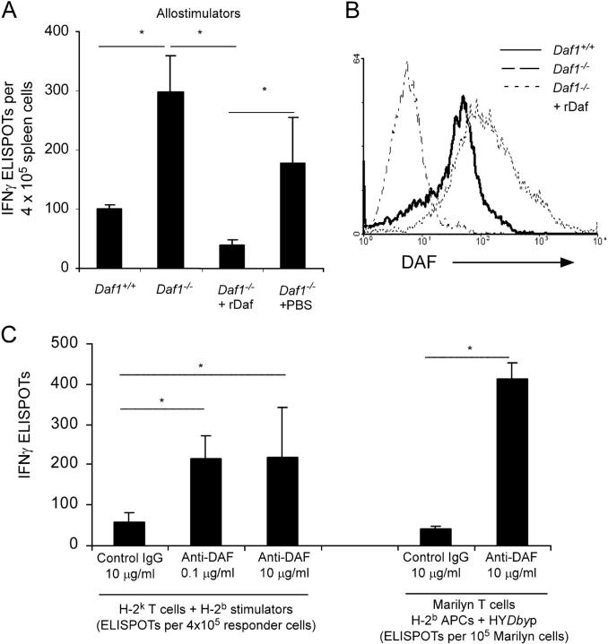
Decay-accelerating factor (Daf) dissociates C3/C5 convertases that assemble on host cells and thereby prevents complement activation on their surfaces. We demonstrate that during primary T cell activation, the absence of Daf on antigen-presenting cells (APCs) and on T cells enhances T cell proliferation and augments the induced frequency of effector cells. The effect is factor D- and, at least in part, C5-dependent, indicating that local alternative pathway activation is essential. We show that cognate T cell-APC interactions are accompanied by rapid production of alternative pathway components and down-regulation of Daf expression. The findings argue that local alternative pathway activation and surface Daf protein function respectively as a costimulator and a negative modulator of T cell immunity and explain previously reported observations linking complement to T cell function. The results could have broad therapeutic implications for disorders in which T cell immunity is important.


Figures

Figure 1.
Figure 1.
Daf deficiency/blockade augments cellular immunity in vitro. (A) IFNγ ELISPOT assays for Daf1 / splenocytes (± reconstitution using lipid-tailed murine rDaf) or Daf1 +/+ splenocytes versus allogeneic H-2 k spleen cells. (B) Flow cytometry of Daf expression levels on cells in A. (C) WT H-2 k T cells stimulated with allogeneic H-2 b spleen cells (left) or Mar TCR transgenic T cells stimulated with HYDby peptide-loaded H-2bAPCs (right) with or without anti-Daf mAb 2C6 or control IgG in IFNγ ELISPOT assays. Means and SD (n = 3–5/group) shown are representative of 2–4 independent experiments; *, P < 0.05.
Figure 2.
Figure 2.
Deficiency of Daf protein augments cellular immunity in vivo. (A) IFNγ ELISPOT assays were performed at rejection of male to female skin grafts (+/+, Daf1 + / +; −/−, Daf1 / ) to assess reactivity to male peptides HYDby (left) and HYUty (right). Each symbol represents an individual animal (mean of triplicate wells, SD < 15%). –, means for all animals per group (n = 4–8); *, P < 0.05 versus Daf1 +/+ to Daf1 +/+. No response (<10 IFNγ producers/500,000 cells) was detected in any animal to control antigens OVA323-339 and β-gal96-103, and no responses were detected in naive mice (not depicted). (B) Spleen cell responses by IFNγ ELISPOT 12 d (top) or 30 d (bottom) after immunization of Daf1 / or Daf1 +/+ mice with HYDby peptide plus CFA. No response was detected to control antigens (not depicted). Means plus SD for three to five animals/group are shown. The results are fully representative of two to four independent experiments. *, P < 0.05.
Figure 3.
Figure 3.
Daf1 / APCs augment T cell immunity in vitro and in vivo via alternative complement activation. (A) H-2 k T cells mixed with allogeneic H-2 b splenic stimulators with or without rDaf were tested in IFNγ ELISPOT assays (means plus SD; n = 3 replicates/group). (B) Proliferation of CFSE-labeled WT H-2 k T cells versus allogeneic Daf1 / or Daf1 +/+ macrophages on d 5. The precursor frequencies of responding T cells were 1.01% (4.1% underwent more than three divisions) against Daf1 / cells versus 0.07% against Daf1 +/+ cells (1.1% underwent more than three divisions). (C) Proliferation of splenic CFSE-labeled H-2 k T cells on day 5 after injection into allogeneic hosts. The mean numbers of transferred cells that underwent more than three cell divisions (n = 3 animals/group) are shown. (D) IFNγ ELISPOT production by Mar T cells stimulated with 10 μM HYDby plus spleen cells. (E) Proliferation of CFSE-labeled Vβ6+ Mar T cells 3 d after adoptive transfer into indicated mice (3 × 10⁶ cells/mouse). (Left) representative plots gated on CFSE+ Vβ6+ cells (inset: % Mar cells/spleen). (Right) quantified means (n = 3 animals/group). (F, left) Daf1 +/+ or Daf1 −/− mice were immunized with OVA323-339 plus anti-C5 mAb or control (1 mg i.p. every other day), and recall assays to OVA323-339 (0.1 μM) were performed by ELISPOT on d 16. *, P < 0.05 versus the response in Daf1 / mice. Right: 3-d in vitro IFNγ ELISPOT assays of H-2 k T cells versus Daf1 +/+ or Daf1 / APCs plus 100 μg/ml anti-C5 mAb or control (top) or H-2 k T cells versus Daf1 +/+ spleen cells plus recombinant C5a (bottom). *, P < 0.05 versus control. Each result is representative of three individual experiments.
Figure 4.
Figure 4.
Daf deficiency on T cells enhances T cell responsiveness. (A) Purified T cells were mixed with WT allogeneic H-2 k splenic stimulator cells in IFNγ ELISPOT assays. (B) T cell–depleted WT Thy1.1 (H-2 b) females were given adoptive transfers of 7.5 × 10⁶ unfractionated, purified splenic T cells isolated from Thy1.2+ Daf1 +/+ or Daf1 / mice. The reconstituted hosts were immunized with HYDby plus CFA, and recall IFNγ ELISPOT assays (means plus SD; n = 3/group) were performed 21 d later. Flow cytometry confirmed that all IFNγ derived from the Thy1.2+ cells (not depicted). *, P < 0.05.
Figure 5.
Figure 5.
T cells and APCs produce complement components during cognate interactions. (A) H-2 k T cells were mixed with allogeneic peritoneal macrophages, flow sorted at predetermined time points, tested for complement expression by quantitative RT-PCR, and compared with unstimulated controls. (B) Electron micrograph of immunostaining using gold–anti-C3 of Mar T cells mixed with female H-2 b peritoneal macrophages plus HYDby peptide (top) or OVA323-339 (bottom). Note gold particles (arrowheads) only in the HYDby peptide-stimulated sample. T, T cell; M, macrophage. Bar, 200 μm. (C) C3, factor B, and factor D in serum-free culture supernates of Mar T cells mixed with HYDby-loaded APCs overnight before (top) and after (middle) cell sorting and reculturing for an additional 3 h. Data are plotted as relative increase over OVA323-339-stimulated controls. C3, factor B, and factor D in serum-free culture supernates of H-2 k T cells stimulated overnight with anti-CD3 (bottom panel), expressed as fold increase over controls. (D) Western blots for factor B (fB, top) and C3 (bottom) in culture supernatants of Mar T cells stimulated with WT or C3 / splenic APCs and HYDby peptide or OVA323-339. Densitometry revealed 20% increase in C3 and 175% increase in factor B with HYDby peptide versus OVA323-339. +, positive control (normal mouse serum); —, negative control (serum from fB / /Crry / or C3−/− mice). E. Plots of Daf expression on macrophages at 0, 24, and 48 h after mixing with Mar T cells plus HYDby peptide or OVA323-339. All results are representative of experiments performed at least two or three times.

References

    1. Janeway, C., Jr., P. Travers, M. Walport, and M.J. Shlomchik. 2004. Innate immunity. In Immunobiology. Garland Publishing, New York. 37–100.
    1. Suresh, M., H. Molina, M.S. Salvato, D. Mastellos, J.D. Lambris, and M. Sandor. 2003. Complement component 3 is required for optimal expansion of CD8 T cells during a systemic viral infection. J. Immunol. 170:788–794. - PubMed
    1. Pratt, J.R., S.A. Basheer, and S.H. Sacks. 2002. Local synthesis of complement component C3 regulates acute renal transplant rejection. Nat. Med. 8:582–587. - PubMed
    1. Nataf, S., S.L. Carroll, R.A. Wetsel, A.J. Szalai, and S.R. Barnum. 2000. Attenuation of experimental autoimmune demyelination in complement-deficient mice. J. Immunol. 165:5867–5873. - PubMed
    1. Kopf, M., B. Abel, A. Gallimore, M. Carroll, and M.F. Bachmann. 2002. Complement component C3 promotes T-cell priming and lung migration to control acute influenza virus infection. Nat. Med. 8:373–378. - PubMed

Publication types

MeSH terms