. 2005 May 16;201(10):1591-602.
doi: 10.1084/jem.20042167. Epub 2005 May 9.

Recruitment of latent pools of high-avidity CD8(+) T cells to the antitumor immune response

Affiliations

Recruitment of latent pools of high-avidity CD8(+) T cells to the antitumor immune response

Anne M Ercolini et al. J Exp Med. .

Abstract

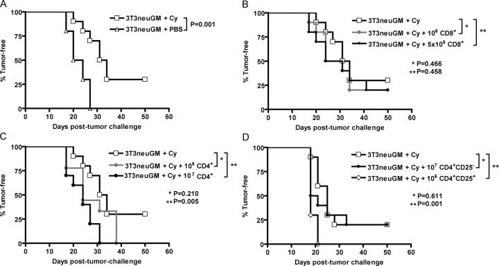
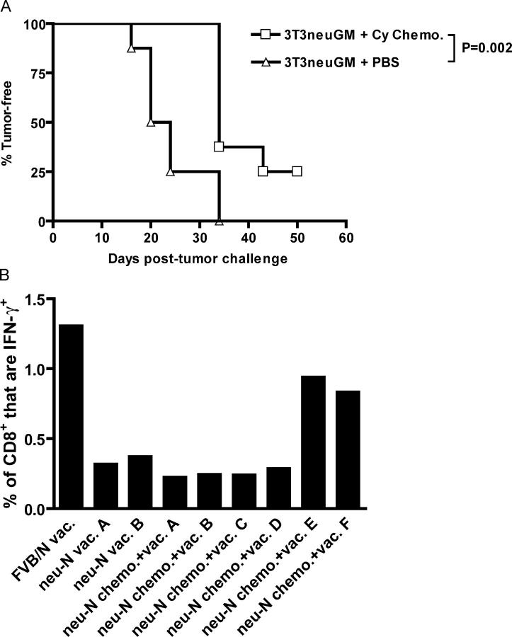
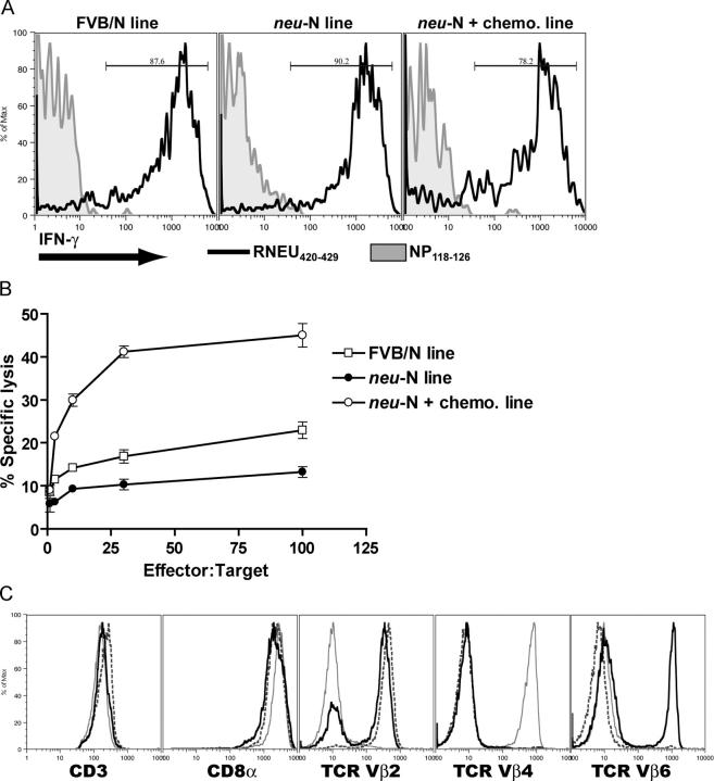
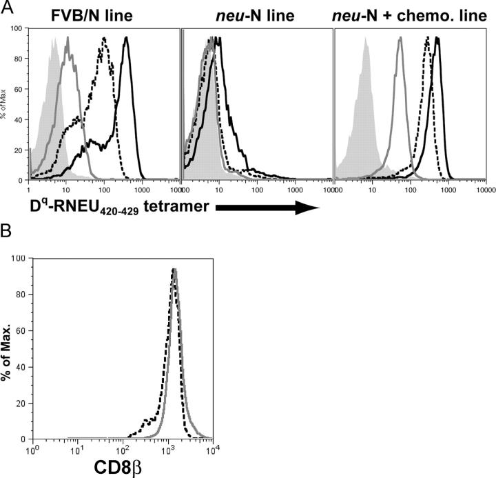
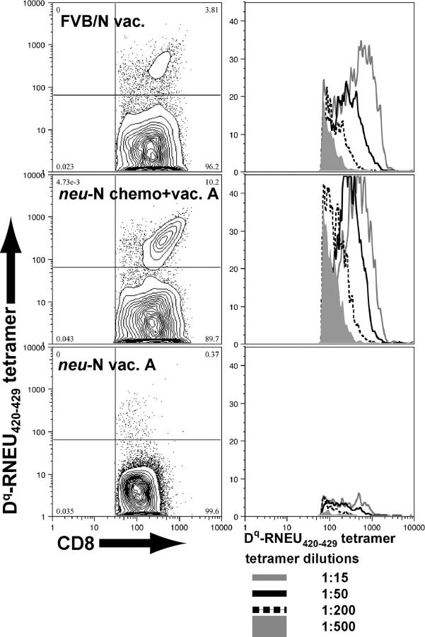
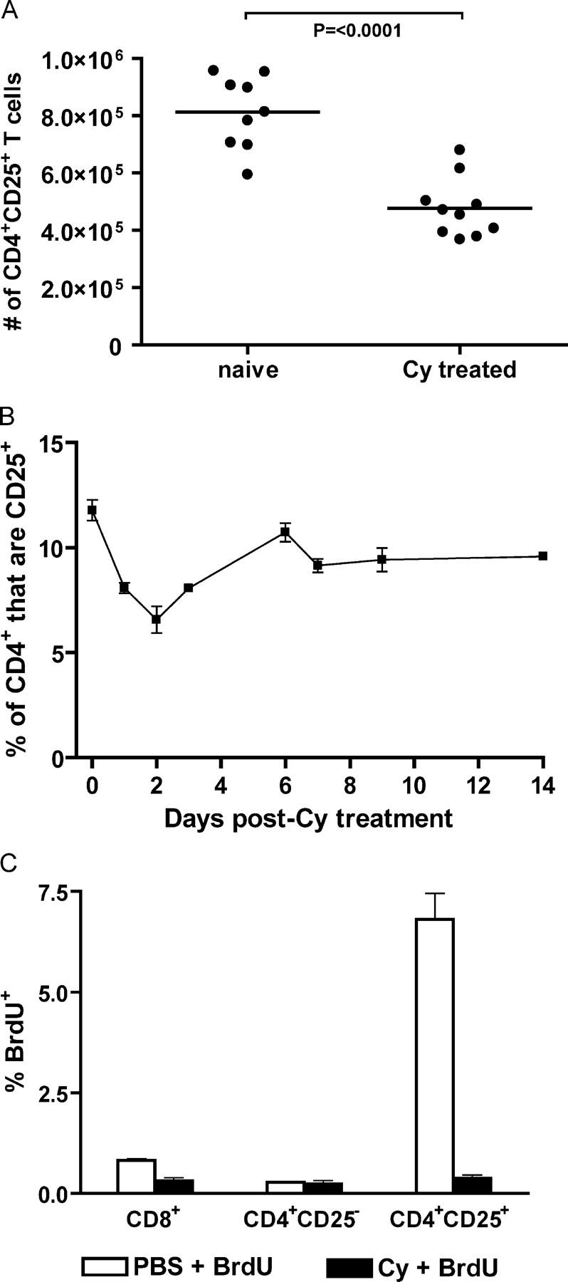
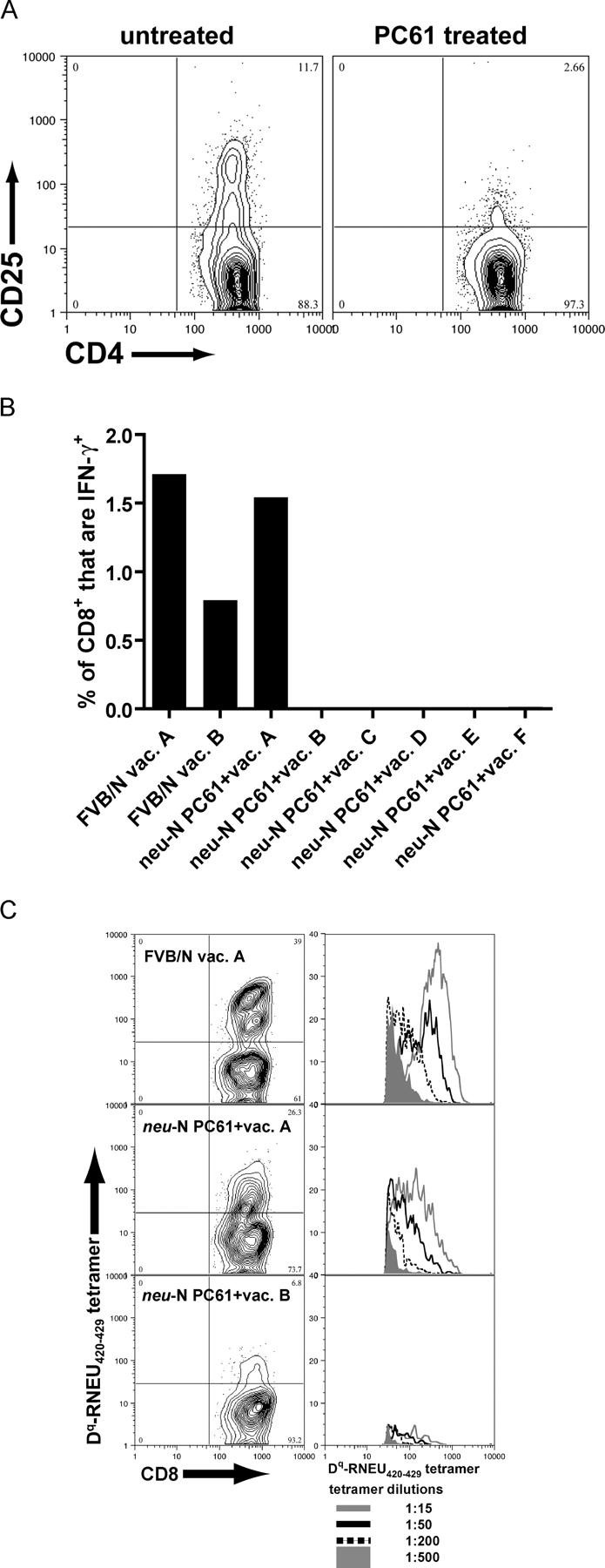
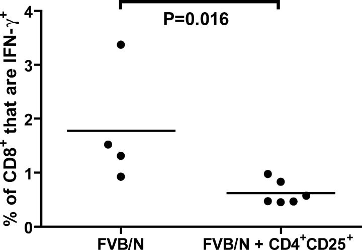
A major barrier to successful antitumor vaccination is tolerance of high-avidity T cells specific to tumor antigens. In keeping with this notion, HER-2/neu (neu)-targeted vaccines, which raise strong CD8(+) T cell responses to a dominant peptide (RNEU(420-429)) in WT FVB/N mice and protect them from a neu-expressing tumor challenge, fail to do so in MMTV-neu (neu-N) transgenic mice. However, treatment of neu-N mice with vaccine and cyclophosphamide-containing chemotherapy resulted in tumor protection in a proportion of mice. This effect was specifically abrogated by the transfer of neu-N-derived CD4(+)CD25(+) T cells. RNEU(420-429)-specific CD8(+) T cells were identified only in neu-N mice given vaccine and cyclophosphamide chemotherapy which rejected tumor challenge. Tetramer-binding studies demonstrated that cyclophosphamide pretreatment allowed the activation of high-avidity RNEU(420-429)-specific CD8(+) T cells comparable to those generated from vaccinated FVB/N mice. Cyclophosphamide seemed to inhibit regulatory T (T reg) cells by selectively depleting the cycling population of CD4(+)CD25(+) T cells in neu-N mice. These findings demonstrate that neu-N mice possess latent pools of high-avidity neu-specific CD8(+) T cells that can be recruited to produce an effective antitumor response if T reg cells are blocked or removed by using approaches such as administration of cyclophosphamide before vaccination.


Figures

Figure 1.
Figure 1.
Adoptive transfer of CD4+CD25+ T reg cells abrogates the immune modulatory effect of cyclophosphamide. 10 neu-N mice/group were vaccinated with either 3T3neuGM alone on day 0 or with cyclophosphamide on day −1 and 3T3neuGM on day 0. Adoptively transferred T cells were given on day 13, as described in Materials and methods. On day 14, all mice were challenged with NT2 tumor cells. Mice were monitored for tumor outgrowth two times/wk. (A) cyclophosphamide given before vaccine results in significant protection (P = 0.001) from NT tumor challenge. Adoptively transferred, purified CD8+ splenocytes (B) were not capable of suppressing the effect of cyclophosphamide, whereas CD4+ splenocytes (C) did significantly inhibit the immune modulatory effects of cyclophosphamide in a dose-dependent fashion (P = 0.005 for 107 CD4+). (D) The experiment was repeated using purified CD4+CD25 splenocytes and purified CD4+CD25+ splenocytes. A significant suppressive effect of the T cells localized to the CD4+CD25+ splenocytes (P = 0.001), and not to the CD4+CD25 splenocytes (P = 0.611). Each study was repeated at least twice with similar results. Cy, cyclophosphamide.
Figure 2.
Figure 2.
RNEU420-429-specific T cells can be identified in polyclonal T cell populations from neu-N mice given vaccine and cyclophosphamide chemotherapy that rejected neu-expressing tumors. (A) Eight neu-N mice/group were tumor challenged on day −3, followed on day 0 by vaccination with or without cyclophosphamide chemotherapy. Mice were monitored for tumor outgrowth two times/wk. All mice that received mock vaccine with or without cyclophosphamide chemotherapy developed tumor by day 35 (not depicted). (B) At the end of the experiment in (A), splenic T cells were isolated, and reactivity to RNEU420-429 was determined by ICS. Plotted is the percentage of CD8+ T cells that secreted IFN-γ in response to RNEU420-429 minus the percentage responding to NP118-126. This experiment was repeated at least three times with similar results. Cy, cyclophosphamide.
Figure 3.
Figure 3.
FVB/N and neu-N–derived CD8+ T cell lines are specific for RNEU420-429 but differentially lyse neu-expressing mammary tumors despite expressing similar levels of cell surface markers. (A) T cell lines were derived from vaccinated FVB/N mice (FVB/N line), neu-N were mice given vaccine alone (neu-N line), or neu-N mice were given vaccine and cyclophosphamide chemotherapy that had rejected an NT tumor challenge (neu-N plus chemotherapy line). IFN-γ ICS was performed after T cells were stimulated overnight with equal numbers of T2Dq cells pulsed with either the irrelevant peptide NP118-126 (shaded histogram) or with RNEU420-429 (black line). (B) CTL assay using the three T cell lines and neu-expressing NT2 tumor targets. These experiments were repeated more than six times with similar results. (C) Cells were stained with antibodies to CD3, CD8α, TCR Vβ2, TCR Vβ4, and TCR Vβ6 as described in Materials and methods. Black line, FVB/N line; dashed line, neu-N line; gray line, neu-N plus chemotherapy line.
Figure 4.
Figure 4.
RNEU420-429–specific CD8+ T cell lines derived from FVB/N and neu-N mice demonstrate quantitatively different avidities for the H-2Dq/RNEU420-429 MHC/peptide complex but no difference in CD8β staining. (A) The three T cell lines were stained with decreasing amounts of H-2Dq/ RNEU420-429 tetramer. Shown is the tetramer staining of gated CD8+ T cells. Black solid line, 1:5 tetramer dilution; dashed line, 1:50 tetramer dilution; gray line, 1:500 tetramer dilution; shaded, no tetramer. (B) T cell lines from neu-N mice given vaccine alone (dashed line) and neu-N mice given cyclophosphamide chemotherapy (gray line) were stained for CD8β on day 6 after stimulation.
Figure 5.
Figure 5.
High-avidity, RNEU420-429-specific T cells can be identified in polyclonal T cell populations from neu-N mice given vaccine and cyclophosphamide chemotherapy that rejected neu-expressing tumors. CD8+ enriched splenocytes were stained with decreasing amounts of H-2Dq-RNEU420-429 tetramer. Three representative samples are shown. Plots on the left are gated on CD8+, CD62Llo lymphocytes and show the 1:50 dilution of tetramer. Corresponding plots on the right show histograms gating on the CD8+, tetramer+ cells stained with decreasing amounts of tetramer.
Figure 6.
Figure 6.
Cyclophosphamide depletes cycling CD4+CD25+ T cells. (A) Axillary LNs from PBS- or cyclophosphamide-treated neu-N mice (10/group) were isolated 48 h after cyclophosphamide administration. Total cell number in each LN was determined, and the number of CD4+CD25+ T cells was calculated based on the percent of CD4+CD25+ T cells determined by flow cytometry. (B) LN cells from neu-N mice (4/group) given cyclophosphamide were isolated and analyzed for the number of CD4+CD25+ T cells on the indicated days. The experiment was repeated three times. (C) Neu-N mice were given cyclophosphamide or PBS on day 0. On day 1, the mice were given a 2-mg dose of BrdU. On day 2, LNs were harvested, and the lymphocytes were stained for CD4, CD8, CD25, and BrdU. This experiment was repeated three times with similar results. Cy, cyclophosphamide.
Figure 7.
Figure 7.
High-avidity RNEU420-429-specific CD8+ T cells are detected after depletion of CD4+CD25+ T reg cells with the CD25+ T cell-depleting antibody, PC61. (A) Depletion was obtained using a single 1-mg dose of PC61 given 4 d before analysis. Splenocytes were isolated and stained with the noncompeting anti-CD25 antibody 7D4. (B) Mice were given 1 mg of PC61 4 d before vaccination. 2 wk after vaccination, splenocytes were isolated, and ICS was performed. Shown are the percent of CD8+ T cells that were RNEU420-429-specific. Similar results have been seen in more than six independent experiments. (C) Splenocytes from (B) were stimulated for 1 wk with RNEU420-429 and then stained with decreasing amounts of H-2Dq/RNEU420-429 tetramer. Three representative samples are shown. Plots on the left are gated on CD8+, CD62Llo lymphocytes and show the 1:50 dilution of tetramer. Corresponding plots on the right show histograms gating on the CD8+, tetramer+ cells stained with decreasing amounts of tetramer.
Figure 8.
Figure 8.
Adoptive transfer of CD4+CD25+ T cells from vaccinated, tumor-bearing neu-N mice suppresses the activation of RNEU420-429-specific CD8+ T cells in FVB/N mice. CD4+CD25+ T cells from vaccinated, tumor-bearing neu-N mice were isolated by cell sorting. 5 × 10⁵ CD4+CD25+ T reg cells were transferred into naive FVB/N mice that were vaccinated the following day. On day 14 after vaccination, splenic CD8+ T cells were isolated, and ICS was performed. Shown are the percent of total CD8+ T cells that produced IFN-γ in response to RNEU420-429. This study was repeated twice with similar results. •, individual mice; solid line, average.

Similar articles

Cited by

References

    1. Sprent, J., and H. Kishimoto. 2002. The thymus and negative selection. Immunol. Rev. 185:126–135. - PubMed
    1. Walker, L.S., and A.K. Abbas. 2002. The enemy within: keeping self-reactive T cells at bay in the periphery. Nat. Rev. Immunol. 2:11–19. - PubMed
    1. O'Garra, A., and P. Vieira. 2004. Regulatory T cells and mechanisms of immune system control. Nat. Med. 10:801–805. - PubMed
    1. Pardoll, D. 2003. Does the immune system see tumors as foreign or self? Annu. Rev. Immunol. 21:807–839. - PubMed
    1. Disis, M.L., and M.A. Cheever. 1997. HER-2/neu protein: a target for antigen-specific immunotherapy of human cancer. Adv. Cancer Res. 71:343–371. - PubMed

Publication types

MeSH terms