. 2005 Jun;14(6):1387-95.
doi: 10.1110/ps.041296305. Epub 2005 May 9.

Novel and economical purification of recombinant proteins: intein-mediated protein purification using in vivo polyhydroxybutyrate (PHB) matrix association

Affiliations

Novel and economical purification of recombinant proteins: intein-mediated protein purification using in vivo polyhydroxybutyrate (PHB) matrix association

Mahmoud Reza Banki et al. Protein Sci. 2005 Jun.

Abstract

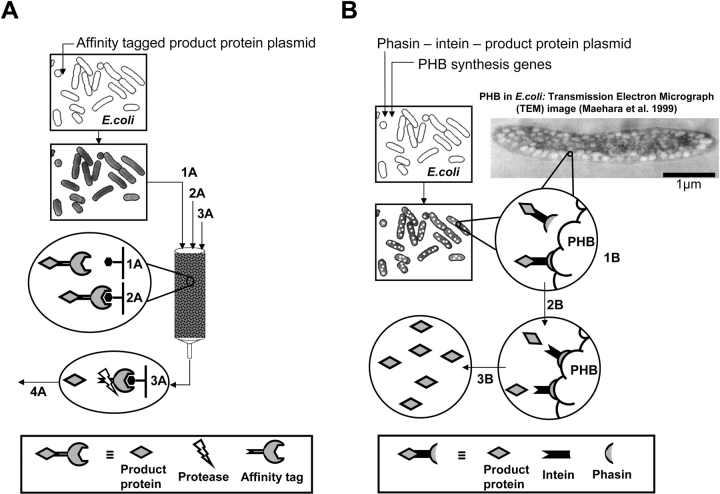
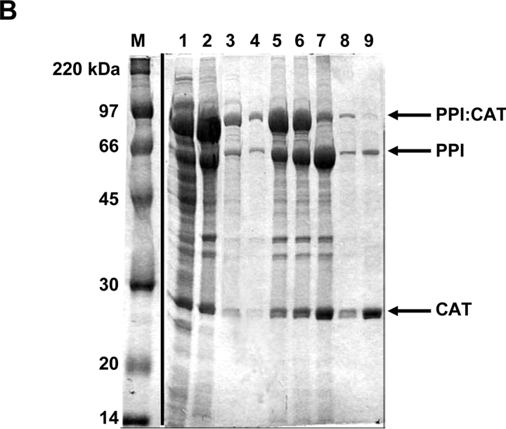
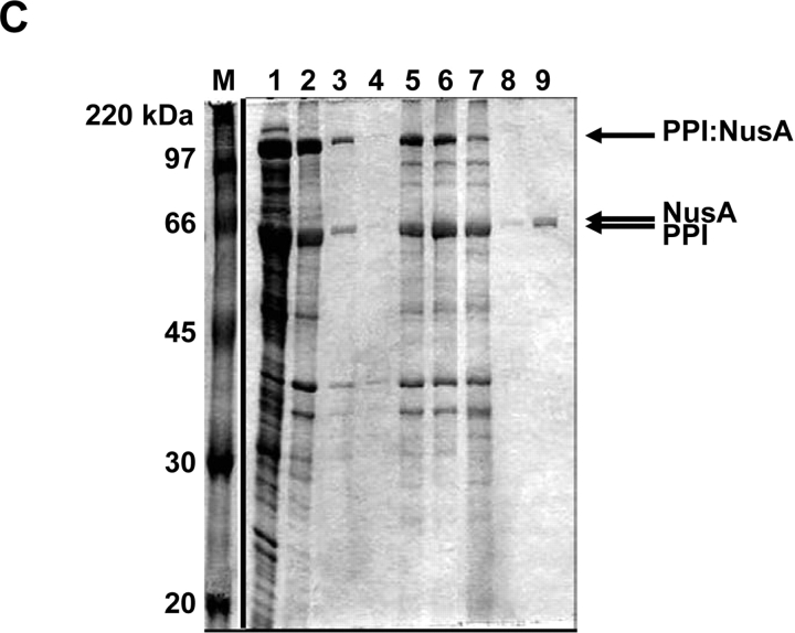
This work combines two well-established technologies to generate a breakthrough in protein production and purification. The first is the production of polyhydroxybutyrate (PHB) granules in engineered strains of Escherichia coli. The second is a recently developed group of self-cleaving affinity tags based on protein splicing elements known as inteins. By combining these technologies with a PHB-specific binding protein, a self-contained protein expression and purification system has been developed. In this system, the PHB-binding protein effectively acts as an affinity tag for desired product proteins. The tagged product proteins are expressed in E. coli strains that also produce intracellular PHB granules, where they bind to the granules via the PHB-binding tag. The granules and attached proteins can then be easily recovered following cell lysis by simple mechanical means. Once purified, the product protein is self-cleaved from the granules and released into solution in a substantially purified form. This system has been successfully used at laboratory scale to purify several active test proteins at reasonable yield. By allowing the bacterial cells to effectively produce both the affinity resin and tagged target protein, the cost associated with the purification of recombinant proteins could be greatly reduced. It is expected that this combination of improved economics and simplicity will constitute a significant breakthrough in both large-scale production of purified proteins and enzymes and high-throughput proteomics studies of peptide libraries.


Figures

Figure 1.
Figure 1.
Comparison of conventional affinity-based protein purification (A) and protein purification using an intein tag and affinity to PHB (B). (A) Conventional affinity-based protein purification: cells containing a plasmid for expression of the affinity tag-product protein fusion are induced and harvested. The cell pellet is resuspended, lysed, and passed over an affinity resin (1A). The column is then washed to rinse away impurities (2A). The fusion is retrieved from the column by addition of excess affinity tag or a displacing substitute and a protease is typically added to cleave off the product protein from the affinity tag (3A). A separation step (4A) salvages the protease and separates the product protein. (B) PHB-intein method of affinity-based protein purification: cells containing two plasmids, one for biosynthesis of PHB granules and another for expression of the phasin-intein tagged product protein, are grown to produce PHB and express the affinity fusion. Harvested cells are lysed and centrifuged to separate soluble components (1B). The insoluble PHB granules with the PHB-bound fusion protein are washed and resuspended in a cleavage-inducing buffer for release of the product protein (2B). A final centrifugation separates the PHB granules and associated proteins from the cleaved product protein, leaving only the product protein in the soluble fraction (3B).
Figure 2.
Figure 2.
Scanning electron micrograph (SEM) images showing PHB granule synthesis in BLR (DE3) and XL1-Blue strains. All samples were grown for 30 h, lysed, dried, and iridium coated. (A) BLR strain carrying pJM9131 (PHB biosynthesis plasmid) grown in LB media. (B) BLR strain carrying a control ampicillin-resistant plasmid grown in lactate-supplemented LB media. (C) BLR strain carrying pJM9131 (PHB biosynthesis plasmid) grown in lactate-supplemented LB media. (D) XL1-Blue strain carrying pJM9131 (PHB biosynthesis plasmid) grown in lactate-supplemented LB media.
Figure 3.
Figure 3.
SDS-PAGE results for phasin affinity to PHB. (A) BLR strain carrying phaP gene (plasmid pET/phaP) induced for 0.5 and 2 h at 37°C. Lane 1, molecular weight marker. Lane 2, preinduction whole-cell lysate. Lanes 3 and 4, soluble fractions of cell lysates at 0.5- and 2-h inductions, respectively. Lanes 5 and 6, insoluble fractions corresponding to lanes 3 and 4. (B) BLR strain carrying the phaP gene (plasmid pET/phaP) and PHB biosynthesis genes (plasmid pJM9131) grown and induced for 8 and 30 h. Lane 1, preinduction whole-cell lysate. Lanes 2 and 3, soluble fractions after 8 and 30 h, respectively. Lanes 4 and 5, insoluble fractions corresponding to lanes 2 and 3. Note the displacement of phasin from the soluble fraction (B, lane 2) to the insoluble fraction (B, lane 5) in the presence of PHB (after 30 h of growth).
Figure 4.
Figure 4.
Maltose binding protein (MBP) purification: BLR strain double transformed with pJM9131 and pET/PPPI:M, grown for 24 h at 37°C in lactate-supplemented media and IPTG-induced for an additional 4 h at the same temperature. Lane 1, supernatant fraction of cell lysate. Lane 2, insoluble fraction of cell lysate. Lanes 3 and 5, decanted wash. Lane 4, molecular weight markers. Lane 6, post-wash pellet. Lanes 710, insoluble fraction for the cleavage time course after 1, 3, 20, and 25 h, respectively. Lanes 1114, soluble fractions corresponding to lanes 710. Lane 15, supernatant from lane 14 after addition of maltose resin and centrifugation.
Figure 5.
Figure 5.
Purification of additional test proteins. (A) β-Galactosidase, (B) chloramphenicol acetyltransferase (CAT), (C) NusA protein. Proteins were expressed and purified as described in text. Lanes: M, molecular weight marker—same for all gels. Lane 1, supernatant fraction of cell lysate. Lane 2, insoluble fraction of cell lysate. Lanes 3 and 4, decanted wash supernatants. Lane 5, post-wash pellet. Lane 6 and 7, insoluble fraction for the cleavage time course after 2 and 30 h, respectively. Lanes 8 and 9, soluble fraction for the cleavage time course after 2 and 30 h, respectively.
Figure 5.
Figure 5.
Purification of additional test proteins. (A) β-Galactosidase, (B) chloramphenicol acetyltransferase (CAT), (C) NusA protein. Proteins were expressed and purified as described in text. Lanes: M, molecular weight marker—same for all gels. Lane 1, supernatant fraction of cell lysate. Lane 2, insoluble fraction of cell lysate. Lanes 3 and 4, decanted wash supernatants. Lane 5, post-wash pellet. Lane 6 and 7, insoluble fraction for the cleavage time course after 2 and 30 h, respectively. Lanes 8 and 9, soluble fraction for the cleavage time course after 2 and 30 h, respectively.

Comment in

  • Proteins from PHB granules.
    Georgiou G, Jeong KJ. Georgiou G, et al. Protein Sci. 2005 Jun;14(6):1385-6. doi: 10.1110/ps.051418305. Protein Sci. 2005. PMID: 15929993 Free PMC article. No abstract available.

References

    1. Ahn, W.S., Park, S.J., and Lee, S.Y. 2000. Production of Poly(3-hydroxybutyrate) by fed-batch culture of recombinant Escherichia coli with a highly concentrated whey solution. Appl. Environ. Microbiol. 66 3624–3627. - PMC - PubMed
    1. Anderson, A.J. and Dawes, E.A. 1990. Occurrence, metabolism, metabolic role, and industrial uses of bacterial polyhydroxyalkanoates. Microbiol. Rev. 54 450–472. - PMC - PubMed
    1. Ausubel, F.M. 1998. Protein analysis. In Current protocols in molecular biology (eds. F.M. Ausubel et al.), Section 10.11.14. John Wiley & Sons, New York.
    1. Chong, S., Mersha, F.B., Comb, D.G., Scott, M.E., Landry, D., Vence, L.M., Perler, F.B., Benner, J., Kucera, R.B., Hirvonen, C.A., et al. 1997. Single-column purification of free recombinant proteins using a self-cleavable affinity tag derived from a protein splicing element. Gene 192 271–281. - PubMed
    1. Doi, Y. 1990. Fermentation and analysis of microbial polyesters. In Microbial polyesters, p. 156. VCH, New York.

Publication types

MeSH terms

LinkOut - more resources