. 2005 May 9;169(3):415-24.
doi: 10.1083/jcb.200411169.

Promotion of importin alpha-mediated nuclear import by the phosphorylation-dependent binding of cargo protein to 14-3-3

Affiliations

Promotion of importin alpha-mediated nuclear import by the phosphorylation-dependent binding of cargo protein to 14-3-3

Christian Faul et al. J Cell Biol. .

Abstract

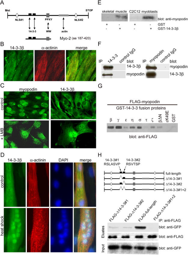
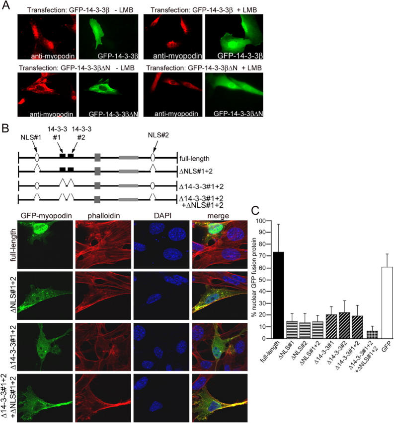
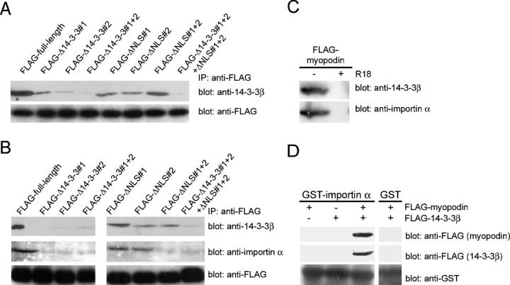
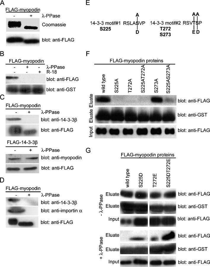
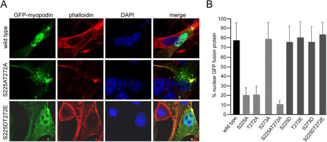
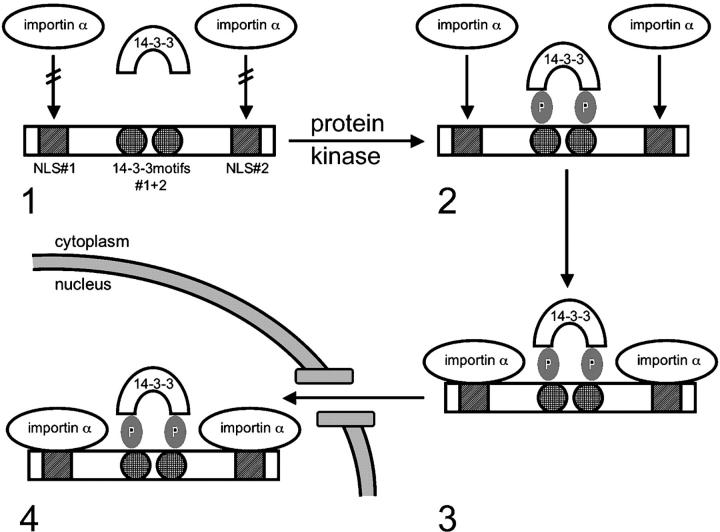
14-3-3 proteins are phosphoserine/threonine-binding proteins that play important roles in many regulatory processes, including intracellular protein targeting. 14-3-3 proteins can anchor target proteins in the cytoplasm and in the nucleus or can mediate their nuclear export. So far, no role for 14-3-3 in mediating nuclear import has been described. There is also mounting evidence that nuclear import is regulated by the phosphorylation of cargo proteins, but the underlying mechanism remains elusive. Myopodin is a dual-compartment, actin-bundling protein that functions as a tumor suppressor in human bladder cancer. In muscle cells, myopodin redistributes between the nucleus and the cytoplasm in a differentiation-dependent and stress-induced fashion. We show that importin alpha binding and the subsequent nuclear import of myopodin are regulated by the serine/threonine phosphorylation-dependent binding of myopodin to 14-3-3. These results establish a novel paradigm for the promotion of nuclear import by 14-3-3 binding. They provide a molecular explanation for the phosphorylation-dependent nuclear import of nuclear localization signal-containing cargo proteins.


Figures

Figure 1.
Figure 1.
Myopodin interacts with 14-3-3 via two consensus 14-3-3–binding motifs. (A) Myopodin contains two NLSs, a PPXY motif, two 14-3-3–binding motifs, and an actin-binding site. Arrows indicate potential interactions with other proteins or with protein domains (WW). The Myo-2 fragment (aa 187–420) was used as bait in a yeast two-hybrid screen. (B) 14-3-3β colocalizes with the Z-disc marker α-actinin in mouse skeletal muscle. (C) In undifferentiated C2C12 myoblasts, myopodin is predominantly found in the nucleus (top left), whereas 14-3-3β is preferentially found in the cytoplasm (top right). Blocking nuclear export with LMB does not affect the nuclear localization of myopodin (bottom left), but leads to the nuclear accumulation of 14-3-3β (bottom right). (D) Heat shock causes nuclear accumulation of 14-3-3β in differentiated myotubes, as visualized by double labeling with DAPI. (E) Myopodin from mouse skeletal muscle (left) and C2C12 myoblast extracts (right) specifically binds to GST–14-3-3β, but not to GST alone. (F) Coimmunoprecipitation experiments show that endogenous myopodin interacts with 14-3-3β in adult mouse heart. (left) IP with anti–14-3-3β; (right) IP with antimyopodin. No binding was found with a control IgG. (G) Myopodin binds to all 14-3-3 isoforms except 14-3-3τ. 14-3-3β lacking the NH2-terminal dimerization domain (βΔN) can bind to myopodin, albeit to a lesser extent. In contrast, 14-3-3ɛ carrying a point mutation (ɛK49E) that is known to abrogate target binding does not interact with myopodin. (H) The two consensus 14-3-3–binding motifs, 14-3-3#1 (sequence RSLASVP) and 14-3-3#2 (sequence RSVTSP), were deleted separately (Δ14-3-3#1 and Δ14-3-3#2) and together (Δ14-3-3#1 + 2). Both FLAG–Δ14-3-3#1 and FLAG–Δ14-3-3#2 display dramatically reduced binding to 14-3-3β as compared with full-length myopodin (full-length FLAG). Deletion of both 14-3-3–binding motifs (FLAG–Δ14-3-3#1 + 2) abrogates binding to 14-3-3β.
Figure 2.
Figure 2.
14-3-3β is required for nuclear import of myopodin. (A) Overexpression of full-length GFP–14-3-3β does not change the nuclear localization of endogenous myopodin (top left), whereas the dominant negative form GFP–14-3-3βΔN causes the nuclear exclusion of myopodin (bottom left). LMB does not reverse the cytoplasmic localization of myopodin that is caused by GFP–14-3-3βΔN (bottom right). (B) Confocal microscopy reveals a predominantly nuclear localization of GFP-tagged, full-length myopodin (top). Deletion of both NLSs (ΔNLS#1 + 2) or both 14-3-3–binding sites (Δ14-3-3#1 + 2) dramatically decreases the nuclear localization of GFP–myopodin. The combined deletion of all four binding motifs (Δ14-3-3#1 + 2 + ΔNLS#1 + 2; bottom) virtually abrogates the nuclear localization of myopodin (bottom). (C) Quantitative analysis is presented as a percentage of nuclear GFP–myopodin. 73.4% of GFP full-length myopodin is found in the nucleus. Deletion of both NLSs separately (ΔNLS#1 and ΔNLS#2) or together (ΔNLS#1 + 2) decreases the amount of nuclear GFP–myopodin to 14.8%, 13.4%, and 14.2%, respectively. Removal of 14-3-3–binding sites (Δ14-3-3#1, Δ14-3-3#2, and Δ14-3-3#1 + 2) reduces nuclear myopodin to 20.0%, 21.8%, and 19.1%, respectively. Combined deletion of all four motifs (Δ14-3-3#1 + 2 + ΔNLS#1 + 2) causes a further reduction of nuclear myopodin to 6.4%. 60.5% of GFP (control) is found in the nucleus. Statistical significance was confirmed by analysis of variance between groups (ANOVA; P < 0.001). Error bars indicate standard deviation.
Figure 3.
Figure 3.
14-3-3 binding to myopodin is required for the interaction between myopodin and importin α. (A) Endogenous 14-3-3β from C2C12 myoblasts coimmmunoprecipitates with myopodin (FLAG-full-length). FLAG–Δ14-3-3#1 and FLAG–Δ14-3-3#2 show impaired binding to 14-3-3β. No binding to 14-3-3β is found for FLAG–Δ14-3-3#1 + 2. In contrast, deletion of one or both NLSs (FLAG-ΔNLS#1, FLAG-ΔNLS#2, and FLAG-ΔNLS#1 + 2) does not interfere with the binding of myopodin to 14-3-3β. Combined deletion of all four motifs (FLAG–Δ14-3-3#1 + 2 + ΔNLS#1 + 2) abrogates the binding of myopodin to 14-3-3β. (B) Binding of 14-3-3β (top) and importin α (middle) to FLAG-tagged myopodin (bottom). Single NLS deletions (FLAG-ΔNLS#1 and FLAG-ΔNLS#2) do not impair the expression of myopodin or the interaction of myopodin with 14-3-3β or importin α. In contrast, the absence of both NLSs (FLAG-ΔNLS#1 + 2) causes loss of importin α binding, whereas 14-3-3β binding is preserved. Deletion of 14-3-3–binding motifs in myopodin (FLAG–Δ14-3-3#1, FLAG–Δ14-3-3#2, and FLAG–Δ14-3-3#1 + 2) abrogates the interaction of myopodin with 14-3-3β and with importin α. (C) The 14-3-3 inhibitory peptide R18 abrogates the binding of myopodin to 14-3-3β (top) and to importin α (bottom). (D) Immobilized GST–importin α, but not GST alone, binds purified FLAG-myopodin in the presence of purified FLAG–14-3-3β. In contrast, no interaction is found between GST–importin α and FLAG–14-3-3β or FLAG-myopodin alone.
Figure 4.
Figure 4.
Two phosphorylated residues in myopodin mediate 14-3-3 binding. (A) SDS-PAGE analysis showing reduced molecular weight of purified FLAG-myopodin after dephosphorylation with λ-PPase. (B) Dephosphorylation of myopodin abrogates binding to GST–14-3-3β. The interaction is also prevented by the 14-3-3–blocking peptide R18. (C) Binding of purified FLAG-myopodin to endogenous 14-3-3β from C2C12 myoblasts is abrogated by dephosphorylation of myopodin (top). In contrast, binding of endogenous myopodin to purified FLAG–14-3-3β is not affected by the dephosphorylation of 14-3-3β (bottom). (D) Dephosphorylation with λ-PPase abrogates the interaction of FLAG-myopodin (bottom) with endogenous 14-3-3β (top) and importin α (middle) from X. laevis extracts. (E) Putative phosphoacceptor sites within the 14-3-3–binding motifs of myopodin. S225 in motif#1, as well as T272 and S273 in motif#2, were substituted with alanine to remove putative phosphorylation sites. Replacement with aspartic acid or glutamic acid was done to mimic phosphorylation. (F) Purified FLAG-tagged, wild-type myopodin interacts with GST–14-3-3β. Substitution of S225 or T272 with alanine (S225A, T272A, and S225AT272A) abrogates 14-3-3β binding. Replacement of S273 with alanine (S273A) does not interfere with the binding of myopodin to 14-3-3β. (G) Substitutions of S225 or T272 with aspartic or glutamic acid, respectively, does not alter the binding of myopodin to 14-3-3 (top). However, after dephosphorylation, only S225DT272E retains strong binding to GST–14-3-3β. Single mutations (S225D and T272E) bind significantly less, and wild-type binding is abrogated.
Figure 5.
Figure 5.
Two phosphorylated residues regulate nuclear localization of myopodin. (A) Confocal imaging of GFP-tagged myopodin in C2C12 myoblasts. Wild-type myopodin shows a predominantly nuclear localization (top), and S225AT272A displays a dramatically decreased nuclear localization (middle). In contrast, S225DT272E shows a primarily nuclear localization (bottom). Rhodamine-labeled phalloidin identifies myopodin-induced actin bundles, and DAPI visualizes nuclei. (B) Quantitative analysis is presented as a percentage of nuclear GFP–myopodin. 77.4% of wild-type myopodin is detected in the nucleus. S225A and T272A show 20.1% and 20.5% nuclear myopodin, respectively. S225AT272A shows a further decrease to 10.5%. In contrast, S273A does not alter nuclear localization (78.8%). S225D, S273D, and T272E show 75.6%, 75.7%, and 80.3% nuclear localization, respectively. S225DT272E displays 83.5% nuclear localization. Statistical significance was confirmed by ANOVA (P < 0.001). Error bars indicate standard deviation.
Figure 6.
Figure 6.
A model for phosphorylation- and 14-3-3-dependent nuclear import of myopodin. (1) When phosphoacceptor sites in 14-3-3–binding motifs#1 (S225) and #2 (T272) are not phosphorylated, myopodin cannot interact with 14-3-3. Therefore, the NLSs in myopodin are not accessible for importin α binding, and myopodin cannot enter the nucleus. (2) After phosphorylation by serine/threonine protein kinases, myopodin binds to 14-3-3, rendering the NLSs accessible for importin α binding. (3) Importin α binds to the NLSs and mediates the nuclear import of myopodin (4).

Similar articles

Cited by

References

    1. Alam, R., N. Hachiya, M. Sakaguchi, S. Kawabata, S. Iwanaga, M. Kitajima, K. Mihara, and T. Omura. 1994. cDNA cloning and characterization of mitochondrial import stimulation factor (MSF) purified from rat liver cytosol. J. Biochem. (Tokyo). 116:416–425. - PubMed
    1. Bihn, E.A., A.L. Paul, S.W. Wang, G.W. Erdos, and R.J. Ferl. 1997. Localization of 14-3-3 proteins in the nuclei of arabidopsis and maize. Plant J. 12:1439–1445. - PubMed
    1. Briggs, L.J., D. Stein, J. Goltz, V.C. Corrigan, A. Efthymiadis, S. Hubner, and D.A. Jans. 1998. The cAMP-dependent protein kinase site (Ser312) enhances dorsal nuclear import through facilitating nuclear localization sequence/importin interaction. J. Biol. Chem. 273:22745–22752. - PubMed
    1. Brunet, A., F. Kanai, J. Stehn, J. Xu, D. Sarbassova, J.V. Frangioni, S.N. Dalal, J.A. DeCaprio, M.E. Greenberg, and M.B. Yaffe. 2002. 14-3-3 transits to the nucleus and participates in dynamic nucleocytoplasmic transport. J. Cell Biol. 156:817–828. - PMC - PubMed
    1. Cahill, C.M., G. Tzivion, N. Nasrin, S. Ogg, J. Dore, G. Ruvkun, and M. Alexander-Bridges. 2001. Phosphatidylinositol 3-kinase signaling inhibits DAF-16 DNA binding and function via 14-3-3-dependent and 14-3-3-independent pathways. J. Biol. Chem. 276:13402–13410. - PubMed

Publication types