. 2005 May 9;169(3):471-9.
doi: 10.1083/jcb.200502122.

Regulation of phototransduction responsiveness and retinal degeneration by a phospholipase D-generated signaling lipid

Affiliations

Regulation of phototransduction responsiveness and retinal degeneration by a phospholipase D-generated signaling lipid

Mary M LaLonde et al. J Cell Biol. .

Abstract

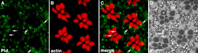
Drosophila melanogaster phototransduction proceeds via a phospholipase C (PLC)-triggered cascade of phosphatidylinositol (PI) lipid modifications, many steps of which remain undefined. We describe the involvement of the lipid phosphatidic acid and the enzyme that generates it, phospholipase D (Pld), in this process. Pld(null) flies exhibit decreased light sensitivity as well as a heightened susceptibility to retinal degeneration. Pld overexpression rescues flies lacking PLC from light-induced, metarhodopsin-mediated degeneration and restores visual signaling in flies lacking the PI transfer protein, which is a key player in the replenishment of the PI 4,5-bisphosphate (PIP2) substrate used by PLC to transduce light stimuli into neurological signals. Altogether, these findings suggest that Pld facilitates phototransduction by maintaining adequate levels of PIP2 and by protecting the visual system from metarhodopsin-induced, low light degeneration.


Figures

Figure 1.
Figure 1.
Phototransduction model. TRP channel activation not only leads to the depolarization of the photoreceptor cell, but it also facilitates the conversion of activated metarhodopsin to inactive rhodopsin via the phosphorylation of Ca2+/CaM kinase, which in turn phosphorylates arrestin2 (Arr2) and causes its dissociation from metarhodopsin (Introduction). Gene names are indicated in italics, phospholipids in bold, and proteins are circled; the circled numbers indicate potential sites of action for Pld as described in the text (see Introduction).
Figure 2.
Figure 2.
Pld null mutant flies display decreased light sensitivity. ERGs were recorded from heterozygous Pld null/CyO (A) and homozygous Pld null (B) adult flies 7 d after eclosion. An intensity–response series to 570-nm stimuli is shown for each fly with intensities of 13.99, 13.38, 12.73, 12.07, and 11.41 log quanta/cm2/s for the control (A) and 15.91, 15.3, 14.65, 13.99, and 13.38 log quant/cm2/s for the Pld null mutant (B). A significant response was not observed for the Pld null mutant at the three lower intensities at which a response was obtained for control flies. Stimulation at 570 nm was chosen because the effects of differences in eye color pigmentation are lower, and on- and off-transients are larger at longer wavelengths (Stark and Wasserman, 1972). Potential was recorded in millivolts as indicated. Three flies were examined for each strain, with similar results observed. (C) The wild-type response was superimposed on the two Pld null mutant ERGs that were conducted within the range examined for the control flies. Light-induced plateau amplitudes were estimated and plotted (D) as a function of the log of the light stimulation intensities to enable a comparison of the wild-type with the Pld null mutant responses. Similar results were obtained when Canton-S was used as the wild-type control.
Figure 3.
Figure 3.
Pld is expressed in the retina and localizes to the photoreceptor cell body. (A–C) Immunostaining of whole-mounted retinas with an affinity-purified, anti-Pld antiserum (A) and with fluorescently labeled phalloidin to visualize the actin-rich rhabdomeres (B). Pld was detected in the photoreceptor cell body and at the base of the rhabdomere (arrows), where a small region of overlap with actin was observed in many cells (C). (D) Retinal tissue sections from Pld null mutant flies raised for 6 wk under a 12-h light/12-h dark cycle were prepared and imaged using electron microscopy. Indistinguishable images were obtained from Canton-S flies raised under identical conditions (not depicted). CB, cell body; R, rhabdomere; SRC, subrhabdomeric cisterna.
Figure 4.
Figure 4.
Pld does not directly transduce light signals, but does morphologically rescue retinal degeneration in the norpA7 mutant. ERGs were obtained from flies raised under continuous light conditions for 1 d: (A) Canton-S, (B) norpA 7, and (C) norpA 7; P{UAS–Pld}/ Rh1. Retinal tissue sections were prepared from flies raised under a 12-h light/12-h dark cycle for 21 d: (D) Canton-S, (E) norpA 7, and (F) norpA 7; P{UAS-Pld}/ Rh1. (D) All 7 photoreceptor cells in all 12 of the complete ommatidia are present in the wild-type Canton-S section, whereas the norpA 7 flies (E) display an irregular ommatidial array that is characterized by intracellular vacuolation (arrows) and missing photoreceptor cells. Only five of the nine complete ommatidia within this section contained seven intact photoreceptor cells. However, a more normal ommatidial structure was observed in norpA 7 flies that overexpressed Pld (F). Although some vacuolation was observed (arrow), all eight complete ommatidia in this section contained seven photoreceptor cells. The experiment was performed three times, and two eyes were sectioned per experimental condition with equivalent results. The entire set of sections was examined, and representative sections were selected for the figures. (G) Western blot analysis of rhodopsin protein levels using a mouse monoclonal antirhodopsin antibody. Tubulin was used as the loading control. Protein was extracted from the following adult heads: Canton-S (lane 1), norpA 7 (lane 2), P{UAS-Pld}/ Rh1 (lane 3), norpA 7; P{UAS-Pld}/ Rh1 (lane 4). Rhodopsin levels were not decreased by Pld overexpression. Sections were representative of three experiments performed. Slightly elevated levels of rhodopsin in the P{UAS-Pld}/ Rh1 sample were observed in this experiment, but this was not a consistent finding based on the other experiments (not depicted).
Figure 4.
Figure 4.
Pld does not directly transduce light signals, but does morphologically rescue retinal degeneration in the norpA7 mutant. ERGs were obtained from flies raised under continuous light conditions for 1 d: (A) Canton-S, (B) norpA 7, and (C) norpA 7; P{UAS–Pld}/ Rh1. Retinal tissue sections were prepared from flies raised under a 12-h light/12-h dark cycle for 21 d: (D) Canton-S, (E) norpA 7, and (F) norpA 7; P{UAS-Pld}/ Rh1. (D) All 7 photoreceptor cells in all 12 of the complete ommatidia are present in the wild-type Canton-S section, whereas the norpA 7 flies (E) display an irregular ommatidial array that is characterized by intracellular vacuolation (arrows) and missing photoreceptor cells. Only five of the nine complete ommatidia within this section contained seven intact photoreceptor cells. However, a more normal ommatidial structure was observed in norpA 7 flies that overexpressed Pld (F). Although some vacuolation was observed (arrow), all eight complete ommatidia in this section contained seven photoreceptor cells. The experiment was performed three times, and two eyes were sectioned per experimental condition with equivalent results. The entire set of sections was examined, and representative sections were selected for the figures. (G) Western blot analysis of rhodopsin protein levels using a mouse monoclonal antirhodopsin antibody. Tubulin was used as the loading control. Protein was extracted from the following adult heads: Canton-S (lane 1), norpA 7 (lane 2), P{UAS-Pld}/ Rh1 (lane 3), norpA 7; P{UAS-Pld}/ Rh1 (lane 4). Rhodopsin levels were not decreased by Pld overexpression. Sections were representative of three experiments performed. Slightly elevated levels of rhodopsin in the P{UAS-Pld}/ Rh1 sample were observed in this experiment, but this was not a consistent finding based on the other experiments (not depicted).
Figure 5.
Figure 5.
Pld overexpression causes activity and light-dependent retinal degeneration. Retinal tissue sections were prepared from flies raised with the following conditions: under a 12-h light/12-h dark cycle for 21 d, (A) +/ Rh1; (B) P{UAS-Pld}/ Rh1; and (D) P{UAS-Pld-H1095N}/ Rh1; in the dark for 21 d, (C) P{UAS-Pld}/ Rh1; or under continuous light for 1 d, (E) P{UAS-Pld}/ Rh1; (F) trp 1; (G) P{UAS-Pld}/ Rh1, trp 1. The bottom panel consists of electron micrographs corresponding to E–G, with R7 labeled. (B) The overexpression of Pld resulted in changes in photoreceptor cell integrity with disarrayed architecture (white circle, outlining a single ommatidium) and widespread intracellular vacuolation (arrow). Only four of the nine complete ommatidia in this section had seven intact photoreceptor cells. The Rh1 promoter drives Pld expression only in R1–6 cells. R7/8 photoreceptors were largely spared, as can be observed in the circled ommatidia or pointed at by the arrow. (C) Maintaining the same flies in the dark substantially decreased the phenotype, as all 10 complete ommatidia in this section contained 7 intact photoreceptor cells, and only limited vacuolation was observed (arrow). (D) No degenerative changes were observed when a catalytically inactive point mutant allele of Pld (H1095N) was overexpressed. (E and F) Retinal disorganization and degeneration was observed in Pld-overexpressing flies after 1 d of continuous light stimulation (only three out of six complete ommatidia retained seven photoreceptor cells; asterisk in EM image in bottom panel shows an example of a degenerating cell; arrow shows that vacuolization and disorganization is also apparent) but not in trp 1 mutant flies. (G) Pld-induced degeneration was suppressed when Pld was overexpressed in the trp-null background, as all seven complete ommatidia maintained seven photoreceptor cells, and vacuolation was not observed (EM image, bottom). Sections are representative of three experiments performed.
Figure 6.
Figure 6.
Pld overexpression restores light responses in the rdgB9 mutant. ERGs were performed on flies raised under continuous light conditions for 1 d: (A) rdgB 9 and (B) rdgB 9; P{UAS-Pld}/ Rh1. The abnormal ERG seen in the rdgB 9 mutant was rescued by the overexpression of Pld, resulting in normal response amplitude with both on- and off-transients. Retinal tissue sections from flies raised under continuous light for 1 or 3 d were prepared and imaged using electron microscopy: (C and F) rdgB 9, (D and G) P{UAS-Pld}/ Rh1, and (E and H) rdgB 9; P{UAS-Pld}/ Rh1. (C) rdgB 9 flies displayed some retinal degeneration 1 d after eclosion. (F) By 3 d after eclosion, more apparent retinal degeneration, with decreased rhabdomere size and increased vacuolation, had ensued. Flies overexpressing Pld had some degeneration 1 d after hatching (D) as described in Fig. 5; but, after 3 d, the flies had even more severe degeneration, with reduced rhabdomeres and degenerating photoreceptor cell bodies (G). In mutant flies overexpressing Pld (E and H), the degeneration caused by Pld expression was reduced. Note that photoreceptor cell R7 (7) appears relatively normal in G and H, which is consistent with the fact that the Rh1 promoter drives expression of Pld only in R1–6. Sections are representative of three experiments performed.
Figure 7.
Figure 7.
Endogenous levels of Pld action protect against retinal degeneration. Retinal tissue sections were prepared from flies raised under continuous light for 1 or 6 d: (A and C) norpA 7 and (B and D) norpA 7; Pldnull. Although the norpA 7 (A) mutant flies had a fairly normal retinal morphology with intact photoreceptor cells and cell bodies soon after eclosion (and the Pld null was indistinguishable from wild type; not depicted), the Pldnull; norpA 7 double mutant (B) already displayed advanced degeneration, with small rhabdomeres and severely reduced cell bodies. No cell bodies were evident except for photoreceptor R7/8. By 6 d after eclosion, degeneration in the norpA 7 (C) mutant was becoming evident (smaller rhabdomeres and some vacuolation); however, by this time, the norpA 7; Pldnulldouble mutant (D) had undergone extensive retinal degeneration, with only rhabdomeres R7/8 remaining apparent in some ommatidia.

Similar articles

Cited by

References

    1. Agam, K., M. von Campenhausen, S. Levy, H.C. Ben-Ami, B. Cook, K. Kirschfeld, and B. Minke. 2000. Metabolic stress reversibly activates the Drosophila light-sensitive channels TRP and TRPL in vivo. J. Neurosci. 20:5748–5755. - PMC - PubMed
    1. Agam, K., S. Frechter, and B. Minke. 2004. Activation of the Drosophila TRP and TRPL channels requires both Ca2+ and protein dephosphorylation. Cell Calcium. 35:87–105. - PubMed
    1. Alloway, P.G., and P.J. Dolph. 1999. A role for the light-dependent phosphorylation of visual arrestin. Proc. Natl. Acad. Sci. USA. 96:6072–6077. - PMC - PubMed
    1. Alloway, P.G., L. Howard, and P.J. Dolph. 2000. The formation of stable rhodopsin-arrestin complexes induces apoptosis and photoreceptor cell degeneration. Neuron. 28:129–138. - PubMed
    1. Boesze-Battaglia, K., O.P. Lamba, A.A. Napoli Jr., S. Sinha, and Y. Guo. 1998. Fusion between retinal rod outer segment membranes and model membranes: a role for photoreceptor peripherin/rds. Biochemistry. 37:9477–9487. - PubMed

Publication types

MeSH terms