Comparative Study
. 2005 May 17;102(20):7263-7.
doi: 10.1073/pnas.0502751102. Epub 2005 May 9.

Coevolution of TCR-MHC interactions: conserved MHC tertiary structure is not sufficient for interactions with the TCR

Affiliations
Comparative Study

Coevolution of TCR-MHC interactions: conserved MHC tertiary structure is not sufficient for interactions with the TCR

Hye-Jung Kim et al. Proc Natl Acad Sci U S A. .

Abstract

The specificity for self-MHC that is necessary for T cell function is a consequence of intrathymic selection during which T cell antigen receptors (TCRs) expressed by immature thymocytes are tested for their affinity for self-peptide:self-MHC. The germ-line-encoded segments of the TCR, however, are believed to have an innate specificity for structural features of MHC molecules. We directly tested this hypothesis by generating a transgenic mouse system in which the protein HLA-DM is expressed at the surface of thymic cortical epithelial cells in the absence of classical MHC molecules. The specialized intracellular function of HLA-DM has removed this MHC class II-like protein from the evolutionary forces that have been hypothesized to shape TCR-MHC interactions. Our study shows that a structural mimic of MHC class II is not sufficient to appropriately interact with the TCRs expressed by developing thymocytes. This result emphasizes the unique complementarity of TCR-MHC interactions that are maintained by the evolutionary pressures dictated by positive selection.


Figures

Fig. 1.
Fig. 1.
Targeting of DM to the cell surface. (A) Tyrosine 248 in the cytoplasmic tail of HLA-DMB was mutated to an alanine to target DM to the cell surface. (B) Cell surface expression of sDM in the mouse B cell line (CH27) transfected with wild-type HLA-DMA and Y248A HLA-DMB. Stably transfected clones were stained for sDM with an antibody against DM (MaP.DM1) or an isotype control (Mags. DO5).
Fig. 2.
Fig. 2.
Naive T cells are not activated by surface DM. LN cells from B6 or B10.BR mice were incubated with CH27 or sDM transfected cells for 72 h, then pulsed with [3H]thymidine. Incorporation of [3H]thymidine was measured 95 h after pulse. The graph shows the level of radioactive thymidine incorporation in B6 LN cells incubated with CH27 (▪) or sDM-transfected CH27 (•) cells. B10.BR LN cells incubated with CH27 (□) or sDM-transfected CH27 (○) cells show a minimal proliferation.
Fig. 3.
Fig. 3.
sDM expression in K14-sDM-transgenic (Tg) mice. (A) Thymic stromal cells prepared from K14-sDM and MHC double knockout mice were stained with antibodies against CDR1 and DM. CDR1+ cells from transgenic-positive (Tg+) and transgenic-negative (Tg-) mice were compared for sDM expression. (B) Thymic stromal cells from K14-sDM and MHC double knockout mice were cultured to enrich for adherent cells. Cells were identified by staining with anti-CD11b for macrophages or the cortical epithelial cell marker DEC205. Cell surface expression of sDM was compared in each population. (C) Detection of HLA-DM in situ. Thymic sections from K14-sDM and MHC double knockout and C57BL/6 mice were immunohistochemically stained for DM. Cortical epithelial cells in K14-sDM mice are stained in red. (Magnification: ×200.)
Fig. 4.
Fig. 4.
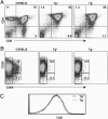
Deficient positive selection of thymocytes in K14-sDM mice. Thymocytes from C57BL/6, K14-sDM, or MHC double knockout mice were stained for CD4, CD8, CD24, CD69, and TCR. (A) CD4 and CD8 staining profile in C57BL/6 (Left), K14-sDM (Center), and MHC double knockout mice (Right). (B) DP cells were gated and analyzed for CD24 and CD69 expression. (C) Expression level of TCR was analyzed in total thymocytes from K14-sDM and MHC double knockout mice.

Similar articles

Cited by

References

    1. Kisielow, P. & von Boehmer, H. (1995) Adv. Immunol. 58, 87-209. - PubMed
    1. Sebzda, E., Mariathasan, S., Ohteki, T., Jones, R., Bachmann, M. F. & Ohashi, P. S. (1999) Annu. Rev. Immunol. 17, 829-874. - PubMed
    1. Ignatowicz, L., Kappler, J. & Marrack, P. (1996) Cell 84, 521-529. - PubMed
    1. Sant'Angelo, D. B., Waterbury, P. G., Cohen, B. E., Martin, W. D., Van Kaer, L., Hayday, A. C. & Janeway, C. A., Jr. (1997) Immunity 7, 517-524. - PubMed
    1. Surh, C. D., Lee, D. S., Fung-Leung, W. P., Karlsson, L. & Sprent, J. (1997) Immunity 7, 209-219. - PubMed

Publication types