

Identification of T-cell epitopes on U1A protein in MRL/lpr mice: double-negative T cells are the major responsive cells
- PMID: 15885135
- PMCID: PMC1782149
- DOI: 10.1111/j.1365-2567.2005.02139.x
Identification of T-cell epitopes on U1A protein in MRL/lpr mice: double-negative T cells are the major responsive cells
Abstract
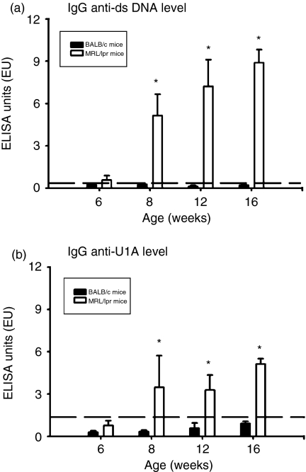
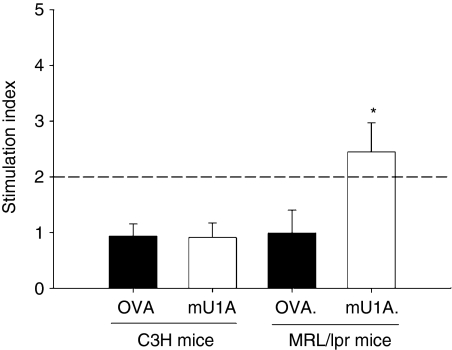
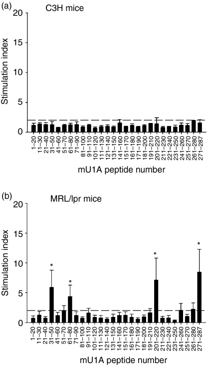
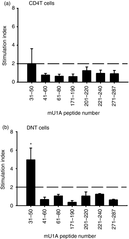
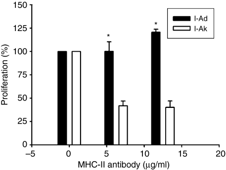
Systemic lupus erythematosus (SLE) is characterized by the existence of a heterogeneous group of autoantibodies such as anti-DNA, chromatin, histone, and ribonucleoprotein antibodies (Abs). Although the B-cell antigenic determinants have been well characterized, very limited data about the T-cell epitopes of self-antigen (Ag) have been reported. In the present study, we analysed auto-T-cell epitopes using bone marrow-derived dendritic cells (BM-DCs) pulsed with murine U1A (mU1A) protein capable of activating autoreactive T cells from unprimed MRL/lpr mice in vitro. The data suggested that there are at least four T-cell epitopes on the U1A protein, U1A31-50, U1A61-80, U1A201-220 and U1A271-287, and U1A31-50 had the most significant T-cell proliferative response. In addition, the main responsive T cells are the CD4- CD8- double-negative subgroup of T cells. Furthermore, we also demonstrated that the activation of double-negative T cells is major histocompatibility complex class II restricted. The study here provides information on T-cell epitope analysis of the U1A antigen using BM-DCs as the effective antigen-presenting cells.
Figures
Similar articles
-
Characterization of self-T-cell response and antigenic determinants of U1A protein with bone marrow-derived dendritic cells in NZB x NZW F1 mice.Immunology. 2001 Jul;103(3):301-9. doi: 10.1046/j.1365-2567.2001.01255.x. Immunology. 2001. PMID: 11454059 Free PMC article.
-
In vivo tolerance breakdown with dendritic cells pulsed with U1A protein in non-autoimmune mice: the induction of a high level of autoantibodies but not renal pathological changes.Immunology. 2002 Jul;106(3):326-35. doi: 10.1046/j.1365-2567.2002.01438.x. Immunology. 2002. PMID: 12100720 Free PMC article.
-
Treatment of murine lupus using nucleosomal T cell epitopes identified by bone marrow-derived dendritic cells.Arthritis Rheum. 2004 Oct;50(10):3250-9. doi: 10.1002/art.20520. Arthritis Rheum. 2004. PMID: 15476240
-
Anti-nucleosome antibodies and T-cell response in systemic lupus erythematosus.Ann Med Interne (Paris). 2002 Dec;153(8):513-9. Ann Med Interne (Paris). 2002. PMID: 12610425 Review.
-
Dysregulated Lymphoid Cell Populations in Mouse Models of Systemic Lupus Erythematosus.Clin Rev Allergy Immunol. 2017 Oct;53(2):181-197. doi: 10.1007/s12016-017-8605-8. Clin Rev Allergy Immunol. 2017. PMID: 28500565 Review.
Cited by
-
Lactobacillus acidophilus Supplementation Exerts a Synergistic Effect on Tacrolimus Efficacy by Modulating Th17/Treg Balance in Lupus-Prone Mice via the SIGNR3 Pathway.Front Immunol. 2021 Dec 10;12:696074. doi: 10.3389/fimmu.2021.696074. eCollection 2021. Front Immunol. 2021. PMID: 34956169 Free PMC article.
-
Baricitinib Attenuates Autoimmune Phenotype and Podocyte Injury in a Murine Model of Systemic Lupus Erythematosus.Front Immunol. 2021 Aug 23;12:704526. doi: 10.3389/fimmu.2021.704526. eCollection 2021. Front Immunol. 2021. PMID: 34497607 Free PMC article.
-
The U1-snRNP complex: structural properties relating to autoimmune pathogenesis in rheumatic diseases.Immunol Rev. 2010 Jan;233(1):126-45. doi: 10.1111/j.0105-2896.2009.00863.x. Immunol Rev. 2010. PMID: 20192997 Free PMC article. Review.
-
Importance of spliceosomal RNP1 motif for intermolecular T-B cell spreading and tolerance restoration in lupus.Arthritis Res Ther. 2007;9(5):R111. doi: 10.1186/ar2317. Arthritis Res Ther. 2007. PMID: 17963484 Free PMC article.
References
-
- Tan EM. Autoantibodies to nuclear antigens (ANA): their immunobiology and medicine. Adv Immunol. 1982;33:167–240. - PubMed
-
- Melissa RA, Arbuckle MR, McClain MT, Rubertone MV, Scofield RH, Dennis GJ, James JA, Harley JB. Development of autoantibodies before the clinical onset of systemic lupus erythematosus. N Engl J Med. 2003;349:1526–33. - PubMed
-
- Fatenejad S, Brooks W, Schwartz A, Craft J. Pattern of anti-small nuclear ribonucleoprotein antibodies in MRL/MP-lpr/lpr mice suggests that the intact U1SnRNP particle is their autoimmunogenic target. J Immunol. 1994;152:5523–31. - PubMed
MeSH terms
Substances
LinkOut - more resources
Full Text Sources
Medical
Research Materials
