Comparative Study
. 2005 May 10;15(9):844-50.
doi: 10.1016/j.cub.2005.03.047.

fsi zebrafish show concordant reversal of laterality of viscera, neuroanatomy, and a subset of behavioral responses

Affiliations
Comparative Study

fsi zebrafish show concordant reversal of laterality of viscera, neuroanatomy, and a subset of behavioral responses

K Anukampa Barth et al. Curr Biol. .

Abstract

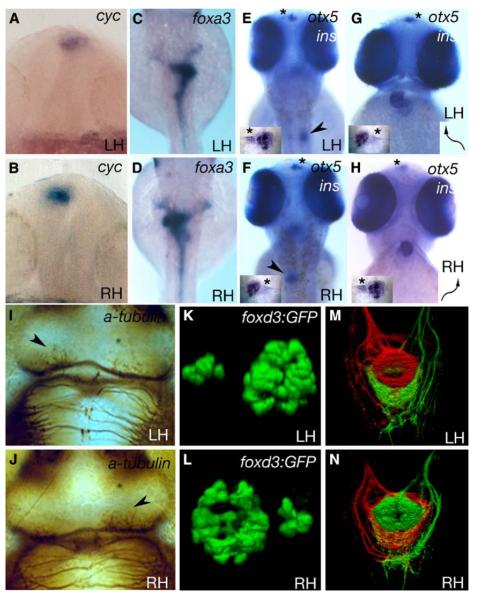
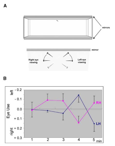
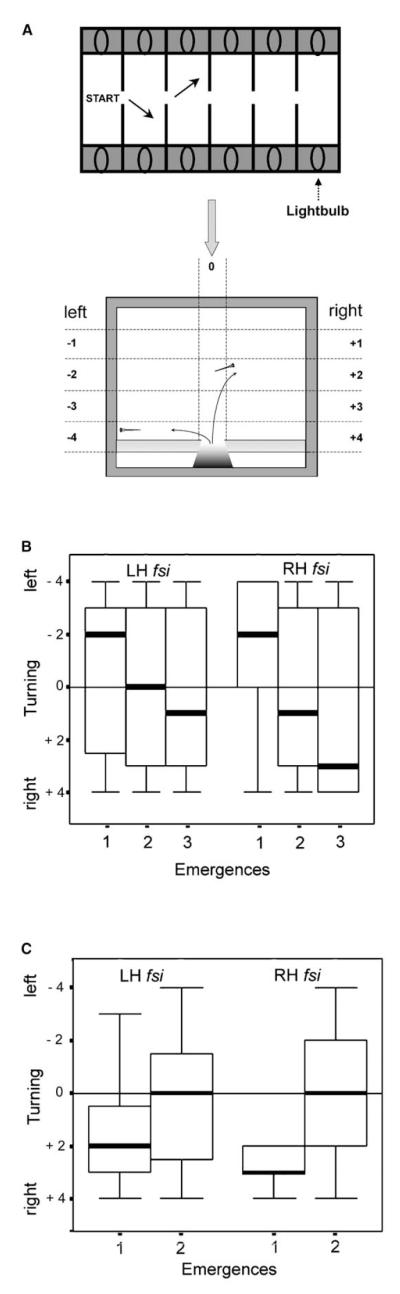
Asymmetries in CNS neuroanatomy are assumed to underlie the widespread cognitive and behavioral asymmetries in vertebrates. Studies in humans have shown that the laterality of some cognitive asymmetries is independent of the laterality of the viscera; discrete mechanisms may therefore regulate visceral and neural lateralization. However, through analysis of visceral, neuroanatomical, and behavioral asymmetries in the frequent-situs-inversus (fsi) line of zebrafish, we show that the principal left-right body asymmetries are coupled to certain brain asymmetries and lateralized behaviors. fsi fish with asymmetry defects show concordant reversal of heart, gut, and neuroanatomical asymmetries in the diencephalon. Moreover, the neuroanatomical reversals in reversed fsi fish correlate with reversal of some behavioral responses in both fry and adult fsi fish. Surprisingly, two behavioral asymmetries do not reverse, suggesting that at least two separable mechanisms must influence functional lateralization in the CNS. Partial reversal of CNS asymmetries may generate new behavioral phenotypes; supporting this idea, reversed fsi fry differ markedly from their normally lateralized siblings in their behavioral response to a novel visual feature. Revealing a link between visceral and brain asymmetry and lateralized behavior, our studies help to explain the complexity of the relationship between the lateralities of visceral and neural asymmetries.


Figures

Figure 1
Figure 1
Some fsi Embryos Show Complete Situs Inversus Frontal (A, B, G, and H) and dorsal (C–F, I–N, and insets in [E]–[H]) views of LH and RH fsi fry. (A and B) Epithalamic expression of cyc. (C and D) Expression of foxa3 in the gut. (E and F) insulin (pancreas, arrowheads in E,F) and otx5 expression (pineal and parapineal; inset, otx5 expression; asterisks indicate parapineal position). (G and H) cmlc2a expression in the heart and otx5 in the pineal and parapineal nuclei (arrows indicate direction of heart looping; inset, otx5 expression; asterisks indicate the position of the parapineal nucleus). Note that the reversed position of the parapineal nucleus in E and G (LH) compared to F and H (RH) is due to different perspectives (dorsal versus frontal views). (I) and (J) show anti-acetylated tubulin labeling of the habenular neuropil (arrowheads indicate the nucleus with more robust labeling). (K and L) 3D reconstructions of pineal (large nucleus) and parapineal (small nucleus) expression of a foxd3:GFP transgene. In all cases, LH and RH fsi fry show opposite laterality of structures. ([A and B] 18–20 somites; [C–J] 2.5 dpf). (M and N) Dorsal view of the intact ventral midbrain in 4 dpf LH and RH fsi fry; shown is a 3D reconstruction of projections from the left (red) and right (green) habenulae within the IPN. There is a DV inversion of projection patterns in the RH fsi fry.
Figure 2
Figure 2
LH and RH fsi Fry Show Opposite Eye Use While Viewing Their Own Reflection for the First Time (A) Schematic representation of the mirror tank and scoring system used for testing. (B) Eye use by LH and RH fsi fry during mirror viewing. For each minute of viewing, scores were calculated as the total duration of right-eye use minus the total duration of left-eye use divided by the combined total of right and left use. The mean of relative eye use and the standard error are shown.
Figure 3
Figure 3
LH and RH fsi Fry Show the Same Directionality Biases When Turning in a Swimway (A) Drawing of swimway tanks (arrows indicate turning movements of fry) and scoring scheme for directionality and angle of turning upon emergence. The turning scores ranged from 1 (shallow turn) to 4 (immediately leaving the medial strip by a sharp turn toward left or right), indicated by horizontal dashed lines. Scores for left turns are negative and for right turns, positive. A score of 0 indicates fry that stayed within the medial strip from entry to the far wall (indicated by the vertical division). The panel was adapted from Watkins et al. [28]. (B) Turning scores in the first three emergences after gradual changes in illumination between chambers shows left-turn bias on first emergence for both LH and RH fsi fry. The box plots show the medians (heavy lines) and distribution across the two quartiles above and below the median representing 50% of the data. Vertical lines extend to the smallest and largest values excluding outliers. (C) Turning scores in the first two emergences after a visual startle stimulus due to a sudden extinction of light in the compartment from which they had emerged and a sudden increase of light in the next compartment. Turning bias is to the right for both groups on first emergence, opposite to that in the no-startle-stimulus condition.
Figure 4
Figure 4
LH and RH fsi Fry Show Different Latencies of Emergence When Confronted with a Conspicuous Vertical Black Stripe Graph showing latency of emergences. The first latency of emergence is similar for both LH and RH fsi fry (and similar to the emergence time in the absence of the black stripe). However, on successive emergences, latencies increase significantly for LH but not for RH fsi fry. Means and standard errors of the mean are shown.

Comment in

Similar articles

Cited by

References

    1. Cooke J. Developmental mechanism and evolutionary origin of vertebrate left/right asymmetries. Biol. Rev. Camb. Philos. Soc. 2004;79:377–407. - PubMed
    1. Malashichev YB, Wassersug RJ. Left and right in the amphibian world: Which way to develop and where to turn? Bioessays. 2004;26:512–522. - PubMed
    1. Vallortigara G, Rogers LJ. Behavioural and Brain Sciences. Cambridge University Press; Cambridge: 2004. Survival with an Asymmetrical Brain: Advantages and Disadvantages of Cerebral Lateralization. - PubMed
    1. Palmer AR. Symmetry breaking and the evolution of development. Science. 2004;306:828–833. - PubMed
    1. Bisgrove BW, Essner JJ, Yost HJ. Multiple pathways in the midline regulate concordant brain, heart and gut left-right asymmetry. Development. 2000;127:3567–3579. - PubMed

Publication types

Substances

LinkOut - more resources