Comparative Study
. 2005 May 10;15(9):862-8.
doi: 10.1016/j.cub.2005.03.050.

Myosin VIIA defects, which underlie the Usher 1B syndrome in humans, lead to deafness in Drosophila

Affiliations
Comparative Study

Myosin VIIA defects, which underlie the Usher 1B syndrome in humans, lead to deafness in Drosophila

Sokol V Todi et al. Curr Biol. .

Abstract

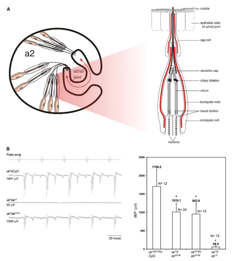
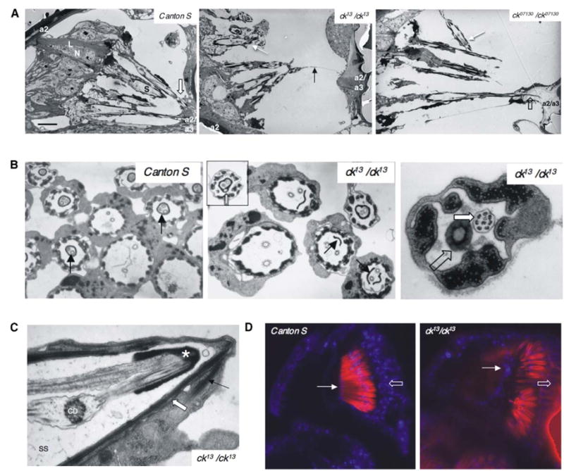
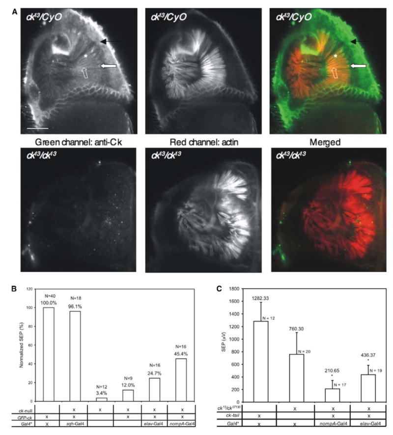
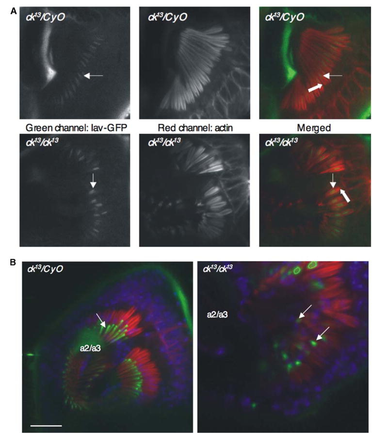
In vertebrates, auditory and vestibular transduction occurs on apical projections (stereocilia) of specialized cells (hair cells). Mutations in myosin VIIA (myoVIIA), an unconventional myosin, lead to deafness and balance anomalies in humans, mice, and zebrafish; individuals are deaf, and stereocilia are disorganized. The exact mechanism through which myoVIIA mutations result in these inner-ear anomalies is unknown. Proposed inner-ear functions for myoVIIA include anchoring transduction channels to the stereocilia membrane, trafficking stereocilia linking components, and anchoring hair cells by associating with adherens junctions. The Drosophila myoVIIA homolog is crinkled (ck). The Drosophila auditory organ, Johnston's organ (JO), is developmentally and functionally related to the vertebrate inner ear. Both derive from modified epithelial cells specified by atonal and spalt homolog expression, and both transduce acoustic mechanical energy (and references therein). Here, we show that loss of ck/myoVIIA function leads to complete deafness in Drosophila by disrupting the integrity of the scolopidia that transduce auditory signals. We demonstrate that ck/myoVIIA functions to organize the auditory organ, that it is functionally required in neuronal and support cells, that it is not required for TRPV channel localization, and that it is not essential for scolopidial-cell-junction integrity.


Figures

Figure 1
Figure 1. JO Function Is Disrupted by ck/myoVIIA Mutations
(A) Diagram of JO. The arrow indicates the direction of a2/a3 movement. The drawing is not to scale. (B) Sound-evoked potentials (SEPs) from the antennal nerve show that ck13/ck07130 flies have lower responses than controls, whereas ck13/ck13 flies have no detectable response. Each trace is the average of ten stimulus/response cycles. Allelic combinations containing ck07130 show SEPs significantly reduced but not absent (p < 0.001), indicating that this is a hypomorphic allele. Error bars indicate the standard deviation.
Figure 2
Figure 2. ck/myoVIIA Mutations Lead to JO Disorganization
(A) ck13 and ck07130 scolopidia, compared to controls (Canton S), show apical detachment (white arrows). In one occasion, out of 14 specimens examined, we observed a residual link (black arrow). The following abbreviations were used: N, neuron; S, scolopale space; L, basal ligament; block arrow, scolopidial attachment to a2/a3 joint; and open block arrow, dendritic cap extending into a2/a3 joint cuticle. The scale bar represents 5 μm. (B) Unlike controls (Canton S, arrow), ck13 shows an incomplete dendritic cap (arrow) and subcompartmentalization (inset, block arrow). When the cap of ck13 completes a profile (open block arrow), it may fail to enclose one of the cilia (block arrow). (C) In detached scolopidia, the cap cell (arrow) detaches from the joint and remains with the scolopidium (block arrow demarcates cell junctions). The following abbreviations were used: SS, scolopale space; CD, ciliary dilation; and asterisk, cap. (D) Nuclear labeling with TO-PRO 3 (blue). a2/a3 joint epithelial cells (arrow) were stained in controls (ck13/CyO) and mutants. Red: actin filaments labeled with Texas Red-Phalloidin; open block arrow: neuronal nuclei. Sections in (D) are equivalent.
Figure 3
Figure 3. ck/myoVIIA Functions in Scolopale Cells and Neurons
(A) Anti-ck/myoVIIA protein antibody labeling endogenous ck/myoVIIA protein in adult ck13/CyO antenna. Rhodamine-phalloidin-stained actin is shown. The arrow indicates neuronal expression; the asterisk, scolopale-cell expression; the block arrow, basal body accumulation; and the open block arrow, apical accumulation. No specific labeling was detected in mutants. Cuticular autofluorescence is observed in both channels. The scale bar represents 20 μm. (B) Full rescue was obtained with sqh-Gal4 (ubiquitous expression). Partial rescue was obtained with nompA-Gal4 (scolopale cells) and elav-Gal4 (neurons). Basal rescue was observed in the absence of any driver, but significantly less than with elav-Gal4 (p < 0.001). (C) myoVIIA/ck tail significantly reduced transduction when expressed in either neurons or scolopale cells (p < 0.001). Error bars indicate standard deviations. For (B) and (C), Gal4*: Gal4 is either elav-Gal4 or nompA-Gal4; no significant difference was found between them. “X” denotes the presence of mutant/construct/driver in genotype.
Figure 4
Figure 4. Localization of Iav-GFP and NompA-GFP in ck/myoVIIA Mutants
(A) Both control (ck13/CyO) and mutant antennae show similar localization of Iav-GFP at the dendritic cilium (arrows) above the location where ck/myoVIIA protein presence is concentrated at the basal body (block arrow). (B) NompA-GFP localization (arrows, green) is unperturbed in ck/myoVIIA antennae in comparison to controls (ck13/CyO), although the cap is fully detached from a2/a3. Blue, TO-PRO3 nuclear labeling; red, actin filaments labeled with Texas Red-phalloidin. The scale bar represents 20 μm.

References

    1. Weil D, Blanchard S, Kaplan J, Guilford P, Gibson F, Walsh J, Mburu P, Varela A, Levilliers J, Weston MD, et al. Defective myosin VIIA gene responsible for Usher syndrome type 1B. Nature. 1995;374:60–61. - PubMed
    1. Gibson F, Walsh J, Mburu P, Varela A, Brown KA, Antonio M, Beisel KW, Steel KP, Brown SD. A type VII myosin encoded by the mouse deafness gene shaker-1. Nature. 1995;374:62–64. - PubMed
    1. Ernest S, Rauch GJ, Haffter P, Geisler R, Petit C, Nicolson T. Mariner is defective in myosin VIIA: A zebra-fish model for human hereditary deafness. Hum Mol Genet. 2000;9:2189–2196. - PubMed
    1. Self T, Mahoney M, Fleming J, Walsh J, Brown SDM, Steel KP. Shaker-1 mutations reveal roles for myosin VIIA in both development and function of cochlear hair cells. Development. 1998;125:557–566. - PubMed
    1. Küssel-Andermann P, El-Amraoui A, Safieddine S, Nouaille S, Perfettini I, Lecuit M, Cossart P, Wolfrum U, Petit C. Vezatin, a novel transmembrane protein, bridges myosin VIIA to the cadherin-catenins complex. EMBO J. 2000;19:6020–6029. - PMC - PubMed

Publication types