

Analysis of NK cells and chemokine receptors in tumor infiltrating CD4 T lymphocytes in human renal carcinomas
- PMID: 15887015
- PMCID: PMC11032824
- DOI: 10.1007/s00262-004-0646-1
Analysis of NK cells and chemokine receptors in tumor infiltrating CD4 T lymphocytes in human renal carcinomas
Abstract
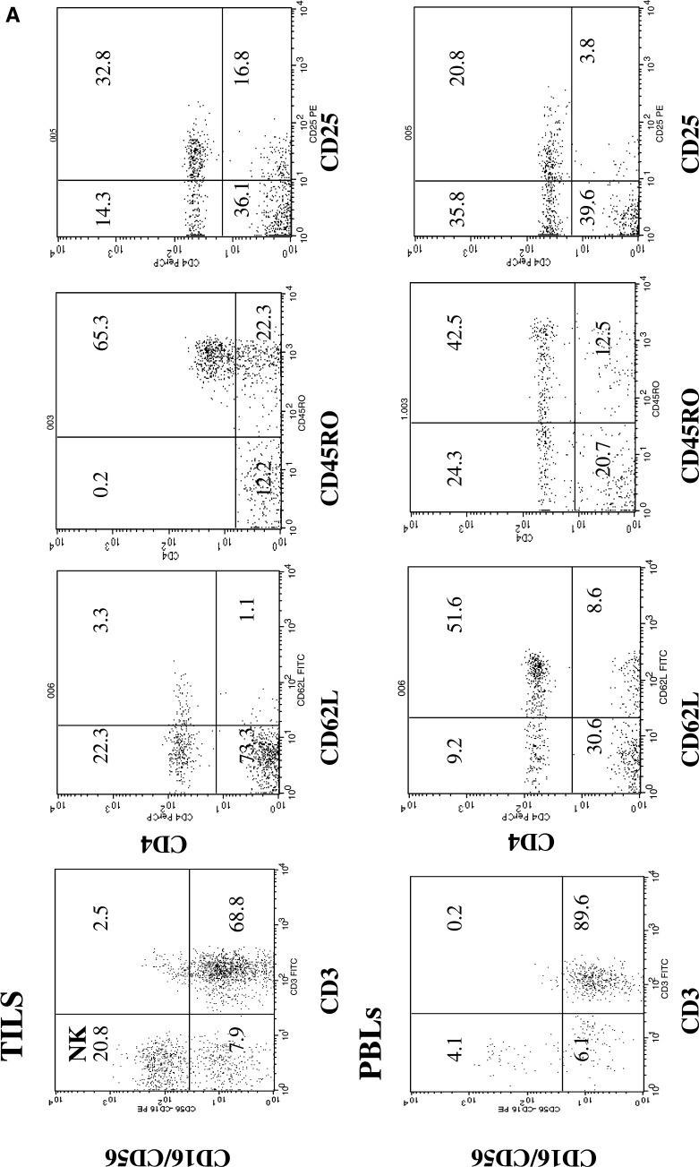
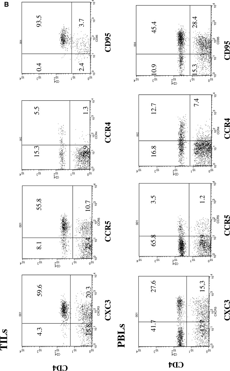
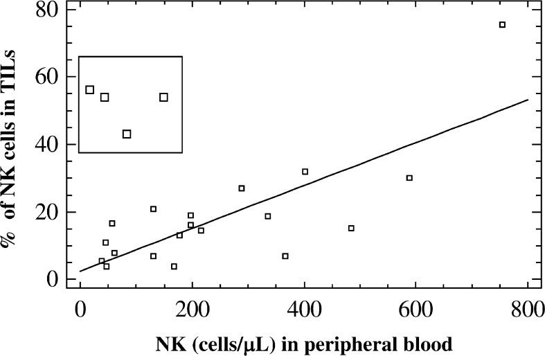
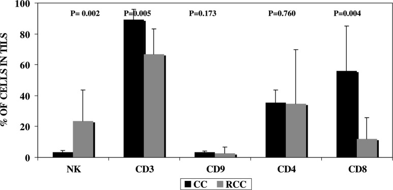
Recent data suggest that chemokines and chemokine receptors mediate leukocyte recruitment of all components of the antitumor response. This study aimed to phenotypically characterize the immune lymphocyte infiltrate in human renal cell carcinomas RCCs and at the invasive margin (tumor-host interface) and to define the association of these findings with established prognostic indicators. Tumor infiltrating lymphocytes TILs were obtained from 24 patients with RCC undergoing radical nephrectomy. Peripheral blood cells from 37 patients were also obtained before surgery. Our findings are consistent with the preferential recruitment of CD4+ Th1-polarized effector memory cells that express CXCR3/CCR5. These cells were the main component of TILs and expressed as CXCR3, CCR5, CD45RO, and CD95. Natural killer (NK) cells were found in significantly higher proportions in TILs of RCCs than in peripheral blood lymphocytes (PBLs) or in other tumors studied (colorectal and breast cancers), where these cells were found in small proportions. No differences in nuclear grade or other studied parameters were observed between the TILs and the lymphocytes present at the invasive margin, which showed a similar composition. However, differences were found according to the tumor stage. First, significantly fewer NK cells were observed in PBLs from metastatic patients. Second, a significantly lower proportion of CCR5/CXCR3/CD4+ cells and a higher proportion of CCR4/CD4+ cells were observed in metastatic patients, suggesting that preferential Th1-polarization may gradually change during the progression of renal cancer cells. Finally, the frequency of CD25/CD4+ cells was higher in metastatic patients. Although the sample of patients with metastasis was small, the overall results suggest a change in composition of the TILs that may potentially confer a selective advantage for tumor growth and may account for the suppression of an effective cytotoxic response.
Figures
References
-
- Bonecchi R, Bianchi G, Bordignon PP, D’Ambrosio D, Lang R, Borsatti A, Sozzani S, Allavena P, Gray PA, Mantovani A, Sinigaglia F. Differential expression of chemokine receptors and chemotactic responsiveness of type 1 T helper cells (Th1s) and Th2s. J Exp Med. 1998;187:129. doi: 10.1084/jem.187.1.129. - DOI - PMC - PubMed
-
- Cabrera CM, Jimenez P, Cabrera T, Esparza C, Ruiz-Cabello F, Garrido F. Total loss of MHC class I in colorectal tumors can be explained by two molecular pathways: beta2-microglobulin inactivation in MSI-positive tumors and LMP7/TAP2 downregulation in MSI-negative tumors. Tissue Antigens. 2003;61:211. doi: 10.1034/j.1399-0039.2003.00020.x. - DOI - PubMed
Publication types
MeSH terms
Substances
LinkOut - more resources
Full Text Sources
Other Literature Sources
Medical
Research Materials
