Comparative Study
. 2005 Apr 22;272(1565):775-84.
doi: 10.1098/rspb.2004.2876.

Molecular preservation in Late Cretaceous sauropod dinosaur eggshells

Affiliations
Comparative Study

Molecular preservation in Late Cretaceous sauropod dinosaur eggshells

M H Schweitzer et al. Proc Biol Sci. .

Abstract

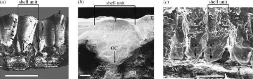
Exceptionally preserved sauropod eggshells discovered in Upper Cretaceous (Campanian) deposits in Patagonia, Argentina, contain skeletal remains and soft tissues of embryonic Titanosaurid dinosaurs. To preserve these labile embryonic remains, the rate of mineral precipitation must have superseded post-mortem degradative processes, resulting in virtually instantaneous mineralization of soft tissues. If so, mineralization may also have been rapid enough to retain fragments of original biomolecules in these specimens. To investigate preservation of biomolecular compounds in these well-preserved sauropod dinosaur eggshells, we applied multiple analytical techniques. Results demonstrate organic compounds and antigenic structures similar to those found in extant eggshells.


Figures

Figure 1
Figure 1
Transmitted light microscopy image of skin fragment from sauropod embryo. Intricate scale pattern is clearly visible.
Figure 2
Figure 2
(a) Tangential ground section of sauropod eggshell, showing external topography in the form of small knobs or tubercles (T), shell units, accretion lines indicating periodic mineral deposition (black arrowheads), and organic cores (OC, arrows) embedded within shell membrane (SM). A pore (P) extends to the outer surface for oxygen exchange. Scale bar, 1 mm. (b) Scanning electron micrograph (SEM) of sauropod shell in tangential section, showing external tubercles (T), shell units, membrane (SM) and organic cores (OC). Scale bar, 200 μm. (c) SEM of extant domestic fowl. Shell units are visible, and organic cores (OC, arrows) are seen embedded in the SM. Scale bar, 100 μm.
Figure 3
Figure 3
Scanning electron micrographs of double-layered (pathological) sauropod eggshell in tangential section. (a) Tubercles (T) delineate the external surface. Preserved shell membrane (M) in between and separating the two layers of shell. Arrow indicates a pore in the depressed region between tubercles connecting exterior to interior of shell. (b) Magnification of area shown in (a), illustrating the difference in texture between the membrane layer (M) and the surrounding shell matrices. Scales are as indicated.
Figure 4
Figure 4
Energy dispersive X-ray (EDX) elemental profile taken through clean fracture of sauropod shell (a) and membrane (b), compared with extant ostrich shell (c) and membrane (d). Element profiles are similar, with a similar content and distribution of trace elements in the membranes not present in the shells. Carbon is higher in the ostrich membrane than the shell, indicating a greater organic content. More labile elements are missing from the fossil analyses (e.g. Cl and Na) as would be expected.
Figure 5
Figure 5
Graphic display of elemental (EDX) data, showing relative values of Ca/C, O/C and O/Ca. These ratios are virtually identical for ostrich and sauropod shell, while ratios obtained from other extant shells vary significantly in these values. Ca/C and O/C values are much higher in sauropod membrane than any extant membrane studied, consistent with a greater degree of mineralization. Extant SMs show greater O/C than Ca/C, a trend also seen in sauropod membrane.
Figure 6
Figure 6
Representative ELISA showing reactivity of pre-immune (white, dark grey bars) and post-immunization (light grey, black) test sera against multiple antigens. Both test sera show significant reactivity with purified ovalbumin, a predominant constituent of extant eggshell matrix and membrane (Nys et al. 2001). Chicken eggshell antibodies react significantly above controls to extracted sauropod and chicken eggshell antigens. Only sauropod antiserum reacts with saruopod shell extracts. No reactivity is demonstrated by any sera with extracted calcite mineral.
Figure 7
Figure 7
Immunohistochemical localization of antigens in ground sections of extant and fossil eggshell. First two columns are imaged using confocal fluorescence microscopy, third column is transmitted light image. (a),(d) Sauropod shell, and (g),(j) chicken shell, incubated with pre-immune sera. (b) Sauropod shell exposed to antisauropod antiserum. (e) Sauropod shell exposed to chicken antiserum. (h) Chicken shell exposed to antisauropod antiserum. (k) Chicken shell exposed to antichicken antiserum. (c),(f),(i),(l) same sample as (b),(e),(h),(k) correspondingly, visualized in transmitted light. Fluorescent label (green) corresponds to location of antibody–antigen complexes. Dark regions show no specific antibody binding. All data were collected under identical exposure and integration conditions. Intensity correlates with degree of antibody binding, pattern of fluorescent distribution corresponds with location of components recognized by antibody. White arrowheads show location of organic cores (OC) within mammillae. CP, arrows show regions of calcite precipitation between mammillae of sauropod shell; ml, mammillary layer of shell; and SM, shell membrane.
Figure 8
Figure 8
Immunohistochemical localization of antigens in ground sections of crocodile eggshell probed with antichicken and antidinosaur shell antibodies. Left panels are imaged using confocal fluorescence microscopy, right panels are transmitted light images. (a) chicken shell antiserum shows strong reactivity throughout the shell matrix, concentrated in the mammillary layer (ml) and the innermost border of the shell membrane (SM). (b) Sauropod shell antiserum shows reduced reactivity of the antiserum to components of crocodile shell matrix, and reactivity is mostly concentrated in the mammillary layer (ml) and the outer SM. (c) Antiserum incubated with excess sauropod extract prior to exposure to crocodile shell to inhibit binding of sauropod-specific antibodies to crocodile epitopes similar in structure to sauropod antigen. See text for discussion.

References

    1. Arias J.L, Fernandez M.S. Role of extracellular matrix molecules in shell formation and structure. World Poult. J. 2001;57:349–355.
    1. Armstrong W.G, Halstead L.B, Reed F.B, Wood L. Fossil proteins in vertebrate calcified tissues. Phil. Trans. R. Soc. B. 1983;301:301–343.
    1. Bada J.L. Amino acid racemization dating of fossils. Annu. Rev. Earth Planet. Sci. 1985;13:241–268.
    1. Baird R.F, Rowley M.J. Preservation of avian collagen in Australian quaternary cave deposits. Paleontology. 1990;33:442–451.
    1. Borja C, Garcia-Pacheco M, Olivares E.G, Scheuenstuhl G, Lowenstein J.M. Immunospecificity of albumin detected in 1.6 million-year-old fossils from Venta Micena in Orce, Granada, Spain. Am. J. Phys. Anthropol. 1997;103:433–441. - PubMed

Publication types