. 2005 Aug;128(Pt 8):1764-77.
doi: 10.1093/brain/awh526. Epub 2005 May 11.

Treatment-responsive limbic encephalitis identified by neuropil antibodies: MRI and PET correlates

Affiliations

Treatment-responsive limbic encephalitis identified by neuropil antibodies: MRI and PET correlates

Beau M Ances et al. Brain. 2005 Aug.

Abstract

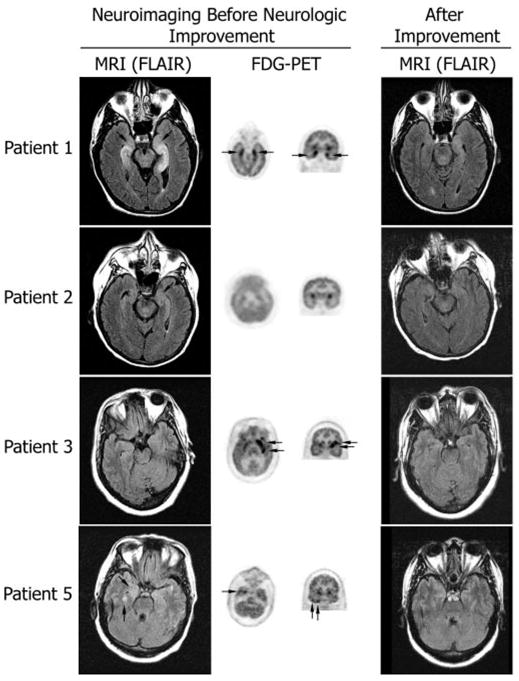
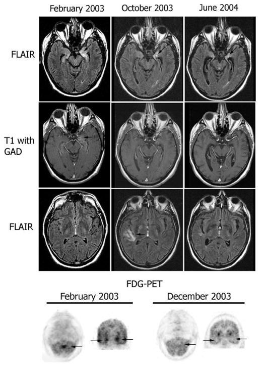
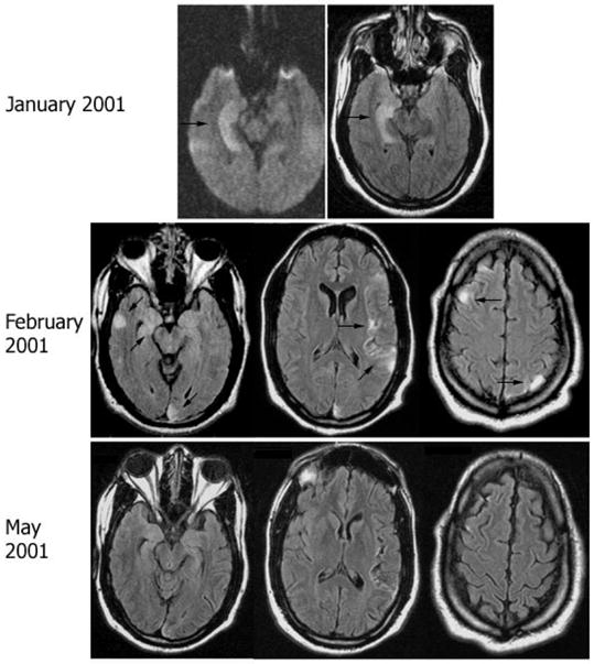
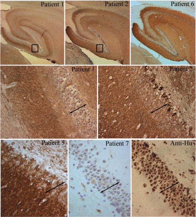
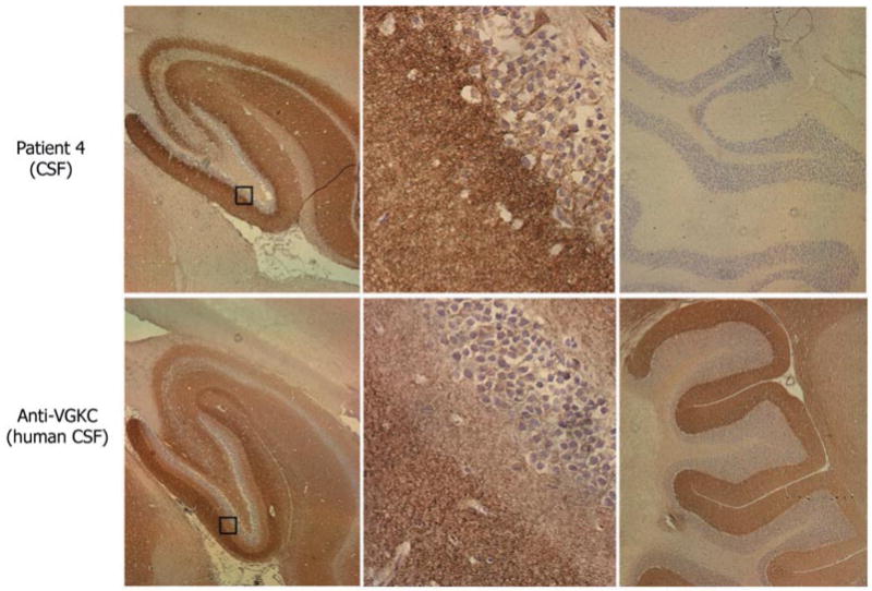
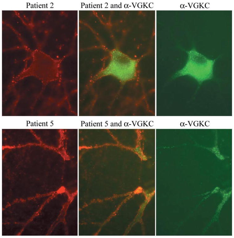
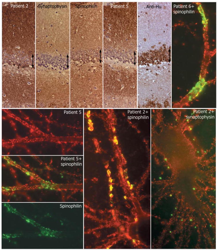
We report seven patients, six from a single institution, who developed subacute limbic encephalitis initially considered of uncertain aetiology. Four patients presented with symptoms of hippocampal dysfunction (i.e. severe short-term memory loss) and three with extensive limbic dysfunction (i.e. confusion, seizures and suspected psychosis). Brain MRI and [(18)F]fluorodeoxyglucose (FDG)-PET complemented each other but did not overlap in 50% of the patients. Combining both tests, all patients had temporal lobe abnormalities, five with additional areas involved. In one patient, FDG hyperactivity in the brainstem that was normal on MRI correlated with central hypoventilation; in another case, hyperactivity in the cerebellum anticipated ataxia. All patients had abnormal CSF: six pleocytosis, six had increased protein concentration, and three of five examined had oligoclonal bands. A tumour was identified and removed in four patients (mediastinal teratoma, thymoma, thymic carcinoma and thyroid cancer) and not treated in one (ovarian teratoma). An immunohistochemical technique that facilitates the detection of antibodies to cell surface or synaptic proteins demonstrated that six patients had antibodies to the neuropil of hippocampus or cerebellum, and one to intraneuronal antigens. Only one of the neuropil antibodies corresponded to voltage-gated potassium channel (VGKC) antibodies; the other five (two with identical specificity) reacted with antigens concentrated in areas of high dendritic density or synaptic-enriched regions of the hippocampus or cerebellum. Preliminary characterization of these antigens indicates that they are diverse and expressed on the neuronal cell membrane and dendrites; they do not co-localize with VGKCs, but partially co-localize with spinophilin. A target autoantigen in one of the patients co-localizes with a cell surface protein involved in hippocampal dendritic development. All patients except the one with antibodies to intracellular antigens had dramatic clinical and neuroimaging responses to immunotherapy or tumour resection; two patients had neurological relapse and improved with immunotherapy. Overall, the phenotype associated with the novel neuropil antibodies includes dominant behavioural and psychiatric symptoms and seizures that often interfere with the evaluation of cognition and memory, and brain MRI or FDG-PET abnormalities less frequently restricted to the medial temporal lobes than in patients with classical paraneoplastic or VGKC antibodies. When compared with patients with VGKC antibodies, patients with these novel antibodies are more likely to have CSF inflammatory abnormalities and systemic tumours (teratoma and thymoma), and they do not develop SIADH-like hyponatraemia. Although most autoantigens await characterization, all share intense expression by the neuropil of hippocampus, with patterns of immunolabelling characteristic enough to suggest the diagnosis of these disorders and predict response to treatment.


Figures

Fig. 1
Fig. 1
MRI and FDG-PET at symptom presentation and MRI after improvement. Axial MRI FLAIR sequences have been matched with axial and coronal FDG-PET to show the areas of maximal involvement. The same MRI region is shown after symptom improvement. Patient 1 had bilateral medial temporal lobe FLAIR abnormalities that correlated with the areas of FDG hyperactivity; the MRI findings are remarkably improved 4 months later. Patient 2 had mild FLAIR abnormalities in the medial temporal lobes with generalized FDG hypoactivity (attributed to antidepressants); the MRI was normal 3 months later. Patient 3 had bilateral non-specific foci of T2 and FLAIR abnormalities in frontal lobes (not shown) and normal temporal lobe findings; the FDG-PET showed hyperactivity in the left frontotemporal region; follow-up MRI 22 months later was unchanged (non-specific abnormalities in frontal lobes; normal temporal lobes) without evolving atrophy. Patient 5 had mild FLAIR abnormalities in the temporal lobes that predominated in the right side, and correlated with the indicated areas of FDG hyperactivity in the right temporal lobe (FDG hyperactivity was also present in the left cerebellum, not shown); the follow-up MRI 27 months later was normal.
Fig. 2
Fig. 2
Follow-up MRI and FDG-PET in patient 7. MRI FLAIR sequences and T1 with gadolinium (T1 with GAD) obtained at three time points during the clinical course of patient 7. Note the presence of bilateral medial temporal lobe FLAIR hyperintensities with progressive evolving atrophy (arrows point to the progressive volume loss in the left hippocampus), and transient FLAIR abnormality in the right insula (October 2003). After gadolinium administration (T1 with GAD), there was mild enhancement in the left medial temporal lobe (February 2003) that resolved in the follow-up studies. The FDG-PET obtained in February 2003 showed hyperactivity in the vermis of the cerebellum (arrow) that preceded by 4 months the development of gait ataxia, and hyperactivity in the medial temporal lobes (bilateral arrows in coronal sections) that correlated with the limbic dysfunction and MRI FLAIR abnormalities in the temporal lobes. The FDG-PET in December 2003 shows resolution of the hyperactivity that correlated with transient stabilization of symptoms and decreased CSF pleocytosis after treatment with cyclophosphamide and corticosteroids.
Fig. 3
Fig. 3
Follow-up MRI in patient 6. MRI FLAIR sequences obtained at different time points of symptom recurrence in patient 6. Note the discrete cortical areas of hyperintensity involving the medial and lateral right temporal region, right frontal lobe, left insular and left parietal and occipital lobes (arrows). Significant improvement was noted in the MRI obtained in May 2001, correlating with clinical improvement of the multifocal encephalitis.
Fig. 4
Fig. 4
Immunohistochemical analysis of neuropil antibodies. Upper row: sagittal sections of rat brain immunoreacted with sera of patients 1, 2 and 6. Note that the three sera show intense reactivity with the neuropil of hippocampus. Patients 2 and 6 harboured the same novel neuropil antibody and abrogated the reactivity of each other in immunocompetition assays; patient 2 had an additional antibody reacting with a subset of neurons in the inner part of the dentate gyrus (shown at high magnification in the middle row). Middle and lower rows show at high magnification the reactivity of sera from patients 1, 2, 5 and 7 and a control anti-Hu. Compare the intracellular reactivity of sera 7 and anti-Hu with the predominant neuropil reactivity of sera 1, 2 and 5 that spare the nuclei and cytoplasm of neurons (except for serum 2 that reacts with a subset of cells). Note that serum 1 has identical reactivity to the serum of a control patient with radioimmunoassay-positive VGKC antibodies shown at low and high magnifications in Fig. 5 (slides mildly counterstained with haematoxylin; upper row ×5; middle and lower rows ×200).
Fig. 5
Fig. 5
Immunohistochemical comparison of the neuropil antibodies of patient 4 with human VGKC antibodies. Sagittal sections of rat brain immunoreacted with CSF of patient 4 and CSF from a control patient with VGKC antibodies determined with the α-dendrotoxin radioimmunoassay. Note the different patterns of hippocampal reactivity (left panels). The neuropil antibody of patient 4 preferentially reacts with the inner aspect of the molecular layer adjacent to the granular cells of the dentate gyrus, while the VGKC antibodies predominantly react with the outer aspect of the molecular layer distant from the granular cells. These differences are better demonstrated in the squares magnified in the middle panels. The neuropil antibody of patient 4 does not react with cerebellum, while VGKC antibodies react with the molecular layer of cerebellum (right panels). Slides mildly counterstained with haematoxylin. Magnification: left panels ×5; middle panels ×400, right panels ×10.
Fig. 6
Fig. 6
Double immunolabelling of hippocampal neuronal cultures with patients’ sera and VGKC antibodies. Using rat hippocampal neurons, the antibodies from patients 2 and 5 (red, left panels) produce widespread labelling of the surface of neurons and dendrites without co-localization (middle panels) with VGKC antibodies (green, right panels). Note that in cultured rat hippocampal neurons, the VGKC antibodies predominantly label the proximal aspect of the neuronal processes and cytoplasm. Sera of 10 other patients with radioimmunoassay-positive VGKC, and monoclonal and polyclonal antibodies to Kv1.2 produced identical reactivity to the VGKC serum used here (data not shown). All panels ×800 (oil).
Fig. 7
Fig. 7
Comparative reactivity of patients’ antibodies with synaptic and dendritic markers. Top row, first five panels: consecutive coronal sections of rat hippocampus reacted with the indicated antibodies. Note the intense and diffuse reactivity of all antibodies (except anti-Hu) with the neuropil of hippocampus, relatively sparing the cell bodies of the neurons of the dentate gyrus (arrows). Sections mildly counterstained with haematoxylin ×400. Other panels: single and double immunolabelling of neuronal cultures with sera of the indicated patients (red) and spinophilin (green) or synaptophysin (green). Note the predominant surface localization of the antigens targeted by all patients’ sera; there is imperfect co-localization (yellow) with the segmental expression of spinophilin, and less frequent co-localization with synaptophysin. All panels ×800 (oil).

Comment in

Similar articles

Cited by

References

    1. Alamowitch S, Graus F, Uchuya M, Reñé R, Bescansa E, Delattre JY. Limbic encephalitis and small cell lung cancer—clinical and immunological features. Brain. 1997;120:923–8. - PubMed
    1. Bataller L, Dalmau J. Paraneoplastic disorders of the central nervous system: update on diagnostic criteria and treatment. Semin Neurol. 2004;24:461–71. - PubMed
    1. Bataller L, Rosenfeld MR, Graus F, Vilchez JJ, Cheung NK, Dalmau J. Autoantigen diversity in the opsoclonus–myoclonus syndrome. Ann Neurol. 2003;53:347–53. - PubMed
    1. Benyahia B, Liblau R, Merle-Beral H, Tourani JM, Dalmau J, Delattre JY. Cell-mediated autoimmunity in paraneoplastic neurological syndromes with anti-Hu antibodies. Ann Neurol. 1999;45:162–7. - PubMed
    1. Bernal F, Graus F, Pifarre A, Saiz A, Benyahia B, Ribalta T. Immunohistochemical analysis of anti-Hu-associated paraneoplastic encephalomyelitis. Acta Neuropathol. 2002;103:509–15. - PubMed

Publication types

MeSH terms