. 2005 Jun;54(6):789-96.
doi: 10.1136/gut.2004.046516.

Production of adiponectin, an anti-inflammatory protein, in mesenteric adipose tissue in Crohn's disease

Affiliations

Production of adiponectin, an anti-inflammatory protein, in mesenteric adipose tissue in Crohn's disease

K Yamamoto et al. Gut. 2005 Jun.

Abstract

Background and aims: A characteristic feature of Crohn's disease (CD) is mesenteric adipose tissue hypertrophy. Mesenteric adipocytes or specific proteins secreted by them may play a role in the pathogenesis of CD. We recently identified adiponectin as an adipocyte specific protein with anti-inflammatory properties. Here we report on expression of adiponectin in mesenteric adipose tissue of CD patients.

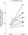
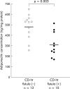
Methods and results: Mesenteric adipose tissue specimens were obtained from patients with CD (n = 22), ulcerative colitis (UC) (n = 8) and, for controls, colon carcinoma patients (n = 28) who underwent intestinal resection. Adiponectin concentrations were determined by enzyme linked immunosorbent assay, and adiponectin mRNA levels were determined by real time quantitative reverse transcription-polymerase chain reaction. Tissue concentrations and release of adiponectin were significantly increased in hypertrophied mesenteric adipose tissue of CD patients compared with normal mesenteric adipose tissue of CD patients (p = 0.002, p = 0.040, respectively), UC patients (p = 0.002, p = 0.003), and controls (p<0.0001, p<0.0001). Adiponectin mRNA levels were significantly higher in hypertrophied mesenteric adipose tissue of CD patients than in paired normal mesenteric adipose tissue from the same subjects (p = 0.024). Adiponectin concentrations in hypertrophied mesenteric adipose tissue of CD patients with an internal fistula were significantly lower than those of CD patients without an internal fistula (p = 0.003).

Conclusions: Our results suggest that adipocytes in hypertrophied mesenteric adipose tissue produce and secrete significant amounts of adiponectin, which could be involved in the regulation of intestinal inflammation associated with CD.


Figures

Figure 1
Figure 1
Haematoxylin-eosin (H&E) staining and immunohistochemical detection of CD68, CD3, and CD20 in hypertrophied mesenteric adipose tissue contiguous with involved intestinal segments of a representative patient with Crohn’s disease (CD). Mesenteric adipose tissue in a normal control subject (A) and hypertrophied mesenteric adipose tissue of a CD patient (B) were stained with H&E. Note that the size of adipocytes in hypertrophied mesenteric adipose tissue is approximately one quarter those of controls (A, B). CD68, CD3, and CD20 expressing cells were stained dark brown (C–E). Many CD68, CD3, and CD20 positive cells were present near the serosa in hypertrophied mesenteric adipose tissue. CD68 positive cells formed a non-caseous epithelioid cell granuloma (C). Nuclear counterstaining: Mayer’s haematoxylin. Original magnification ×200.
Figure 2
Figure 2
Immunohistochemical staining of adiponectin in the mesenteric adipose tissue of a representative control and Crohn’s disease (CD) patient. The mesenteric adipose tissue of the control (B) and the hypertrophied mesenteric adipose tissue of a CD patient (D) reacted with antiadiponectin antibody. No reaction product was detected against non-immune serum in the mesenteric adipose tissue of the control (A) and hypertrophied mesenteric adipose tissue of the CD patient (C). Adiponectin staining appeared as a red fluorescence rim in the periphery of the cytoplasm of adipocytes (B, D). Nuclear counterstaining: 4′, 6-diamidino-2-phenylindole. Original magnification: ×200.
Figure 3
Figure 3
Adiponectin concentrations in mesenteric adipose tissue. Adiponectin concentrations were determined by enzyme linked immunosorbent assay (ELISA) and values expressed in ng of adiponectin per mg of total homogenate protein. (A) Data are individual and mean values (horizontal bar) of adiponectin concentrations in normal mesenteric adipose tissues of controls (C), ulcerative colitis patients (UC), Crohn’s disease patients (CD-n), and hypertrophied mesenteric adipose tissues of CD patients (CD-ht). (B) Data are individual and mean values (horizontal bars) of adiponectin concentrations in normal mesenteric adipose tissues of CD patients (CD-n) and hypertrophied mesenteric adipose tissues of CD patients (CD-ht) from the same patients.
Figure 4
Figure 4
Adiponectin release from the mesenteric adipose tissue in short term culture. Release of adiponectin from adipose tissue was determined by enzyme linked immunosorbent assay and values expressed in ng of adiponectin per ml of conditioned media. Data are individual and mean values (horizontal bars) of adiponectin release from normal mesenteric adipose tissues of controls (C), ulcerative colitis patients (UC), Crohn’s disease patients (CD-n), and hypertrophied mesenteric adipose tissues of CD patients (CD-ht).
Figure 5
Figure 5
Quantitative real time reverse transcription-polymerase chain reaction analysis of adiponectin mRNA expression in mesenteric adipose tissue. Data are individual and mean values (horizontal bars) of adiponectin cDNA molecules per molecule of β-actin cDNA in normal mesenteric adipose tissues of Crohn’s disease patients (CD-n) and hypertrophied mesenteric adipose tissues of CD patients (CD-ht) from the same patients.
Figure 6
Figure 6
Correlation between adiponectin concentrations in hypertrophied mesenteric adipose tissue of Crohn’s disease patients and leucocyte count (A), haemoglobin level (B), platelet count (C), and serum C reactive protein (CRP) levels (D). These laboratory data were determined just prior to surgery.
Figure 7
Figure 7
(A) Interleukin 6 (IL-6) concentrations in the mesenteric adipose tissue of controls (C) and hypertrophied mesenteric adipose tissue of Crohn’s disease patients (CD-ht). IL-6 concentrations were determined by enzyme linked immunosorbent assay and values expressed in pg of IL-6 per mg of total homogenate protein. (B) Correlation between adiponectin and IL-6 concentrations in hypertrophied mesenteric adipose tissue of CD patients.
Figure 8
Figure 8
Adiponectin concentrations in hypertrophied mesenteric adipose tissue of Crohn’s disease (CD) patients with or without internal fistula. Data are individual and mean values (horizontal bars) of adiponectin concentrations in hypertrophied mesenteric adipose tissues of CD patients without an internal fistula (CD-ht fistula (−)) and those of CD patients with an internal fistula (CD-ht fistula (+)).
Figure 9
Figure 9
Relationship between body mass index and tissue concentrations (A) or release (B) of adiponectin in controls, ulcerative colitis patients (UC), Crohn’s disease patients (CD-n), and hypertrophied mesenteric adipose tissue of CD patients (CD-ht).

Comment in

References

    1. Sheehan AL, Warren BF, Gear MW, et al. Fat-wrapping in Crohn’s disease: pathological basis and relevance to surgical practice. Br J Surg 1992;79:955–8. - PubMed
    1. Smedh K, Olaison G, Nystrom PO, et al. Intraoperative enteroscopy in Crohn’s disease. Br J Surg 1993;80:897–900. - PubMed
    1. Weakley FL, Turnbull RB. Recognition of regional ileitis in the operating room. Dis Colon Rectum 1971;14:17–23. - PubMed
    1. Fazio VW, Jones IT. Standard surgical treatment of Crohn’s disease of the small intestine and ileocolitis. In: Lee ECG, Nolan DJ, eds. Surgery of inflammatory bowel disorders. Edinburgh: Churchill Livingstone, 1987:147–56.
    1. Borley NR, Mortensen NJ, Jewell DP, et al. The relationship between inflammatory and serosal connective tissue changes in ileal Crohn’s disease: evidence for a possible causative link. J Pathol 2000;190:196–202. - PubMed

MeSH terms