. 2005 Jun;54(6):835-42.
doi: 10.1136/gut.2004.043034.

Nuclear factor {kappa}B inactivation in the rat liver ameliorates short term total warm ischaemia/reperfusion injury

Affiliations

Nuclear factor {kappa}B inactivation in the rat liver ameliorates short term total warm ischaemia/reperfusion injury

H Suetsugu et al. Gut. 2005 Jun.

Abstract

Background: In hepatic ischaemia/reperfusion injury, activated liver macrophages (Kupffer cells) are dominantly regulated by a transcription factor, nuclear factor kappaB (NFkappaB), with respect to expression of inflammatory cytokines, acute phase response proteins, and cell adhesion molecules.

Aims: We assessed whether inactivation of NFkappaB in the liver could attenuate total hepatic warm ischaemia/reperfusion injury.

Methods: We studied rats with hepatic overexpression of inhibitor kappaBalpha super-repressor (IkappaBalpha SR) caused by a transgene introduced using an adenoviral vector. Hepatic ischaemia/reperfusion injury was induced under warm conditions by total occlusion of hepatoduodenal ligament structures for 20 minutes, followed by reperfusion. Controls included uninfected and control virus (AdLacZ) infected rats.

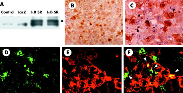
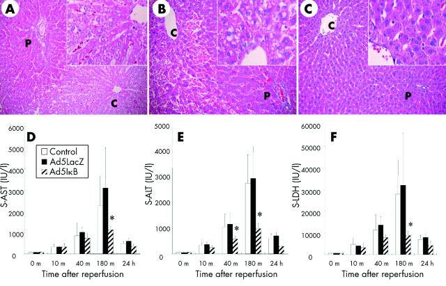
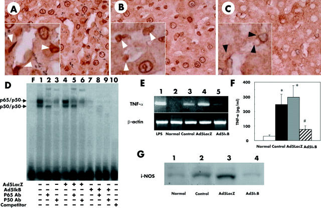
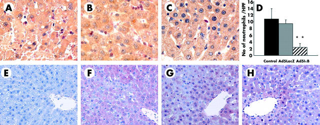
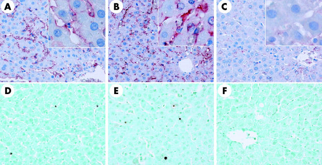
Results: IkappaBalpha SR was overexpressed in Kupffer cells as well as in hepatocytes, blocking nuclear translocation of NFkappaB (p65) into the nucleus after reperfusion. Gene transfection with IkappaBalpha SR, but not with LacZ, markedly attenuated ischaemia/reperfusion injury, suppressing inducible nitric oxide synthase and nitrotyrosine expression in the liver. Moreover, no remarkable hepatocyte apoptosis was detected under IkappaBalpha SR overexpression.

Conclusions: Adenoviral transfer of the IkappaBalpha SR gene in the liver ameliorates short term warm ischaemia/reperfusion injury, possibly through attenuation of hepatic macrophage activation.


Figures

Figure 1
Figure 1
Efficiency of adenovirus mediated gene transfer. (A) Extracts from liver homogenate (20 μg) from uninfected, Ad5LacZ infected, and Ad5IκB infected animals were examined by western blot against inhibitor κBα (IκBα). Black arrow indicates haemagglutinin (HA) tagged IκBα super-repressor (IκB SR). (B, C) Immunohistochemistry of HA tagged IκBα SR (B ×200, C ×400). Black arrowheads indicate HA positive non-parenchymal cells in the liver. Immunofluorescent staining of surface antigen of macrophages (ED-1; D) and β-galactosidase (E), and their superimposed image (F) are presented (×400). Each datum is representative of four separate experiments.
Figure 2
Figure 2
Histological analysis of the liver and liver function tests after ischaemia/reperfusion. Haematoxylin-eosin staining on liver tissues from uninfected (A), Ad5LacZ infected (B), and Ad5IκB infected (C) rats 180 minutes after ischaemia/reperfusion. Necrosis in hepatocytes around central veins and infiltration of inflammatory cells, which were observed in uninfected and Ad5LacZ infected rats, were almost totally blocked in Ad5IκB infected animals. Each picture is representative of six individual animals in each group. Serum (s) aspartate aminotransferase (AST) (D), alanine aminotransferase (ALT) (E), and lactate dehydrogenase (LDH) (F) levels were measured in uninfected, Ad5LacZ infected, and Ad5IκB infected rats at each time point indicated (n = 6 respectively). After reperfusion, the increase in AST, ALT, and LDH in Ad5IκB infected animals was significantly lower than those in uninfected and Ad5LacZ infected animals at the indicated times (*p<0.05).
Figure 3
Figure 3
Uninfected (A), Ad5LacZ infected (B), and Ad5IκB infected (C) livers with ischaemia/reperfusion (×400) underwent immunohistochemical staining of nuclear factor κB (NFκB) p65. After reperfusion, a large number of nuclei were stained with the antibody in uninfected (A) and Ad5LacZ infected (B) livers. In Ad5IκB induced livers, only a small number of positive nuclei were detected (C). High power views showed that a large number of nuclei in non-parenchymal liver cells (NPCs, white arrowheads) as well as in hepatocytes were positive for p65 in uninfected (A, inset) and Ad5LacZ infected (B, inset) livers. In contrast, in the Ad5IκB infected liver, the nuclei of approximately 85% of hepatocytes were negative for p65 staining and only about 15% of NPCs were positive for p65 (C, inset). Black arrowheads indicate nucleus of NPCs, which were negative for p65. (D) Gel shift assay for NFκB. Nuclear extracts from fresh liver samples were assayed for NFκB DNA binding activity by electrophoretic mobility shift assay using a radiolabelled consensus NFκB site probe, as described in materials and methods. A 50-fold cold competitor was added to lane 10. Antibodies against p65 (RelA) or p50 were used for the supershift assay, respectively (α-p65 lanes 2, 5, and 8; α-p50 lanes 3, 6, and 9). Data are representative of three separate experiments. (E) Messenger RNA expression of tumour necrosis factor α (TNF-α). After reperfusion, expression of TNF-α mRNA was strongly induced in uninfected and Ad5LacZ infected animals (lanes 3 and 4). In Ad5IκB infected animals (lane 5), expression of TNF-α mRNA was remarkably suppressed. Results are representative of three separate experiments. cDNA taken from rat liver injected with lipopolysaccharide (LPS) (10 mg/kg) for two hours was used as a positive control (lane 1). (F) Serum concentration of TNF-α was also determined in each animal by means of ELISA at three hours after reperfusion. (G) Western blot for inducible nitric oxide synthase (iNOS) protein. After reperfusion, increased amounts of iNOS were observed in uninfected (lane 2) and Ad5LacZ infected (lane 3) animals. In Ad5IκB infected animals (lane 4), the amount of iNOS was significantly smaller than in the other groups. Results are representative of three separate experiments. Normal, untreated normal rat liver.
Figure 4
Figure 4
Infiltration of neutrophils was estimated by means of naphthol AS-D chloroacetate esterase staining in uninfected (A), Ad5LacZ infected (B), and Ad5IκB infected (C) livers. (D) Number of esterase positive polymorphonuclear cells was counted in 10 high power fields (×400) in each animal; **p<0.01. (E–H) Immnunohistochemistry of 4-hydroxy-2′-nonenal (HNE) adducts in the liver. HNE adducts were not detected in untreated normal rat livers (E) while significant amounts of HNE adducts were detected after reperfusion in uninfected (F), Ad5LacZ infected (G), and Ad5IκB infected (H) livers, particularly surrounding central veins (×200). Each picture is representative of six individual animals in each group.
Figure 5
Figure 5
Immnunohistochemistry of nitrotyrosine (A–C) and single strand DNA (ssDNA) (D–F) in the liver. Nitrotyrosine adducts were detected preferentially along the sinusoidal area, but not in hepatocytes, after short term ischaemia/reperfusion in uninfected (A) and Ad5LacZ infected (B) livers. This increase in nitrotyrosine expression along sinusoids was dramatically suppressed in Ad5IκB infected livers (C) (×200). A relatively small number of ssDNA containing nuclei were detected along the sinusoidal area at three hours after reperfusion in uninfected (D) and Ad5LacZ infected (E) livers, while occasionally such nuclei were also detected among hepatocytes. Gene transfer of IκBα SR suppressed the appearance of ssDNA containing cells after reperfusion (F) (×200). Each picture is representative of six individual animals in each group.

References

    1. Sugawara Y, Kubota K, Ogura T, et al. Increased nitric oxide production in the liver in the perioperative period of partial hepatectomy with Pringle’s maneuver. J Hepatol 1998;28:212–20. - PubMed
    1. Caldwell-Kenkel JC, Currin RT, Tanaka Y, et al. Reperfusion injury to endothelial cells following cold ischemic storage of rat livers. Hepatology 1989;10:292–9. - PubMed
    1. Jaeschke H, Farhood A. Neutrophil and Kupffer cell-induced oxidant stress and ischemia-reperfusion injury in rat liver. Am J Physiol 1991;260:G355–62. - PubMed
    1. Lentsch AB, Kato A, Yoshidome H, et al. Inflammatory mechanisms and therapeutic strategies for warm hepatic ischemia/reperfusion injury. Hepatology 2000;32:169–73. - PubMed
    1. Liu P, McGuire GM, Fisher MA, et al. Activation of Kupffer cells and neutrophils for reactive oxygen formation is responsible for endotoxin-enhanced liver injury after hepatic ischemia. Shock 1995;3:56–62. - PubMed

Publication types

MeSH terms