. 2005 Jun 1;24(11):2043-52.
doi: 10.1038/sj.emboj.7600676. Epub 2005 May 12.

Formation of a novel surface structure encoded by Salmonella Pathogenicity Island 2

Affiliations

Formation of a novel surface structure encoded by Salmonella Pathogenicity Island 2

Dipshikha Chakravortty et al. EMBO J. .

Abstract

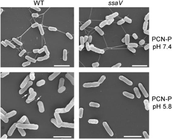
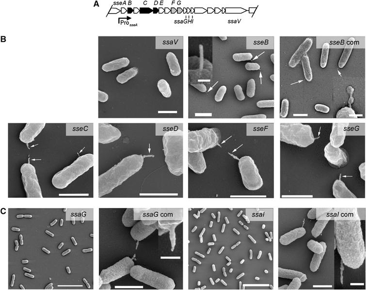
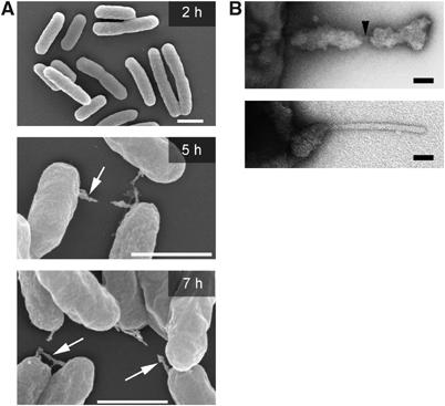
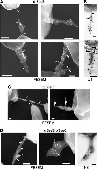
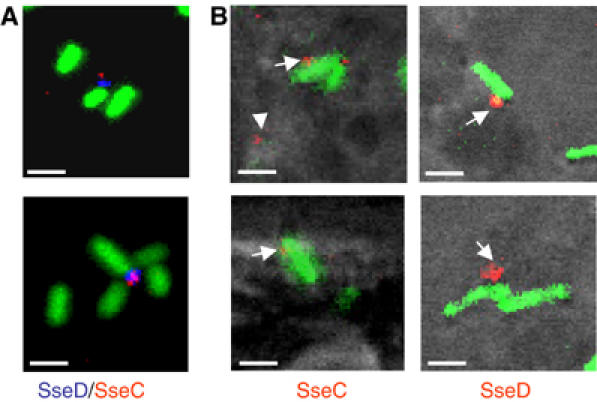
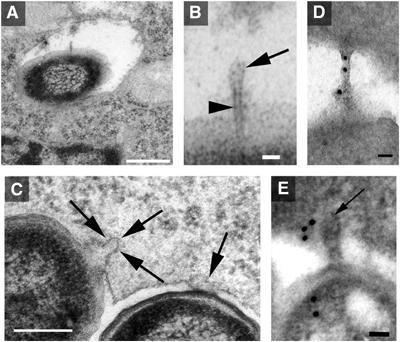
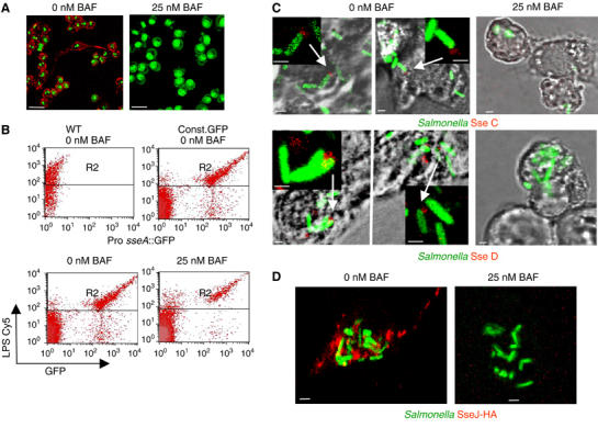
The type III secretion system (T3SS) encoded by Salmonella Pathogenicity Island 2 (SPI2) is essential for virulence and intracellular proliferation of Salmonella enterica. We have previously identified SPI2-encoded proteins that are secreted and function as a translocon for the injection of effector proteins. Here, we describe the formation of a novel SPI2-dependent appendage structure in vitro as well as on the surface of bacteria that reside inside a vacuole of infected host cells. In contrast to the T3SS of other pathogens, the translocon encoded by SPI2 is only present singly or in few copies at one pole of the bacterial cell. Under in vitro conditions, appendages are composed of a filamentous needle-like structure with a diameter of 10 nm that was sheathed with secreted protein. The formation of the appendage in vitro is dependent on acidic media conditions. We analyzed SPI2-encoded appendages in infected cells and observed that acidic vacuolar pH was not required for induction of SPI2 gene expression, but was essential for the assembly of these structures and their function as translocon for delivery of effector proteins.


Figures

Figure 1
Figure 1
SPI2-dependent formation of surface structures in vitro. S. Typhimurium WT and a mutant strain deficient in the SPI2-encoded T3SS (ssaV) were grown in minimal media that induced expression of SPI2 (PCN-P, pH 7.4) or in minimal media inducing SPI2 expression and secretion of substrate proteins (PCN-P, pH 5.8). The bacteria were grown for 16 h with aeration, and aliquots of the cultures were taken and prepared for FESEM analyzes. The appearance of SPI2-dependent polar appendages is indicated by arrows. Similar appendages were observed for S. Typhimurium WT grown in low magnesium minimal media at pH 5.0 (data not shown). Scale bars represent 2 μM.
Figure 2
Figure 2
Effect of mutations in genes encoding T3SS components and secreted proteins on the formation of appendages. (A) Genetic organization of SPI2 genes investigated in this study. Genes encoding translocon subunits (SseB, C, D) and translocated effectors (SseF and G) are indicated by black and gray symbols, respectively. (B) Strains deficient in a structural component of the T3SS (ssaV) or in various translocon or effector proteins were grown in PCN-P media at pH 5.8 and analyzed by FESEM. Appendages were missing on the ssaV strain. Note the appearance of regular needle-like structures for the sseB strain (inset shows magnified view) and of irregular appendages for the other mutant strains. Plasmid complementation of the sseB strain was performed (sseB com). Scale bars represent 1 μm, 100 nm (inset sseB) and 250 nm (inset sseB com). (C) Mutant strains deficient in ssaG or ssaI, and mutant strains harboring a plasmid expressing ssaGHI for complementation (com) were analyzed. Appendage formation was also absent in an ssaH mutant strain (not shown), but was not restored in the complemented strains. Scale bars represent 5 μm for overview images and for the complemented strains 1 μm and 250 nm in the inset.
Figure 3
Figure 3
(A) Kinetics of formation of SPI2-dependent surface structures. S. Typhimurium wild type was cultured overnight in PCN media at pH 7.4. Bacteria were harvested by centrifugation and used for inoculation of PCN-P media at pH 5.8. Incubation at 37°C was continued with aeration and aliquots of the culture were taken at various time points for FESEM analysis. Appendages are indicated by arrows. Scale bars represent 2 μm. (B) Higher magnification view of negatively-stained single appendages revealing the inner cylindrical needle-like structure (arrow) and different extent of proteinaceous sheaths. Bacteria were cultured for 9 h. Scale bars correspond to 50 nm (upper panel) and 25 nm (lower panel).
Figure 4
Figure 4
Detection of SPI-2-encoded proteins by immunoelectron microscopy. S. Typhimurium wild type was grown overnight in PCN-P media at pH 5.8 and cells were processed for immuno-EM. Fixed bacteria were incubated with antisera against SseB (A) or SseC (C) and protein A-coated gold-particles with a diameter of 15 nm. The distribution of the gold-particles (white dots) is detectable over the sheathed parts of the appendage (arrows). Both antibodies did not label the inner cylindrical structures of the appendages (arrowheads). (B) Immunogold labeling of SseB in ultrathin sections (UT). Note that the unlabeled inner structure (arrowhead) spans the entire cell envelope (CM, cytoplasmic membrane; OM, outer membrane). Since the bacteria were pre-embedded, immunogold labeling of SseB (dark dots) is only detectable on the bacterial exterior. (D) For double labeling, antibodies against SseB (arrowheads) and SseC (arrows) were detected with protein A-coated gold-particles of 10 nm for SseB and 15 nm for SseC. A negative-stained (NS) sample of a double-labeled bacterium is shown. Scale bars represent 100 nm.
Figure 5
Figure 5
Detection of SPI2-encoded translocon proteins in vitro and in vivo using immunofluorescence. (A) S. Typhimurium WT harboring pFPV25.1 for the constitutive expression of GFP was grown in PCN-P at pH 5.8 for 7 h. Bacteria were harvested and processed for immunostaining. SseD was detected after incubation with rabbit antiserum against recombinant SseD followed by incubation with a Cy5-labeled secondary antibody (blue). SseC was detected by incubation with rabbit antiserum directly labeled with TRITC (red). (B) RAW macrophages were infected with Salmonella WT harboring pFPV25.1 12 h after infection, cells were fixed and immunostained for SseC and SseD (red). Note the appearance of SseC and SseD on the surface of intracellular bacteria (arrows), and also distant to the bacteria (arrowheads). Scale bars represent 1 μm.
Figure 6
Figure 6
Ultrathin section analysis of intracellular Salmonella. RAW264.7 macrophages were infectedz with S. Typhimurium WT, and 7 h after infection, cells were fixed, dehydrated and embedded in epoxy resin. Analysis of ultrathin sections revealed the expression of appendages that were distant to the phagosomal membrane (A, B) or connecting the bacterial envelope to the phagosomal membrane (CE). Depicted in (B) is a structure that exhibits a needle-like inner core (arrowhead) and a sheathed tip of the appendage (arrow). Immunogold labeling for SseB was performed (D, E). Scale bars represent 500 μm (A), 250 nm (C), 50 nm (D, E) and 25 nm (B).
Figure 7
Figure 7
Secretion and translocation of SPI2 proteins requires acidic phagosomal pH. (A) Phagosome acidification is inhibited by low concentrations of BAF. Various concentrations of BAF (5–100 nM), or an equal amount of the solvent DMSO were added to RAW macrophages. After incubation for 1 h, AO was added to a final concentration of 1 μM and the fluorescence of living cells was analyzed by confocal microscopy. Red fluorescence indicating acidic vesicles was absent at BAF concentrations of 10 nM or higher (only shown for 25 nM BAF, for further details, see Supplementary Figure 4). Scale bars represent 16 μm. (B) Effect of BAF on expression of an SPI2 reporter gene by intracellular S. Typhimurium. RAW macrophages were infected with S. Typhimurium WT without plasmid or harboring plasmid pLS824 (Pro sseA::GFP fusion) or pFVP25.1 (constitutive GFP expression). After infection, 25 nM BAF was added and maintained throughout the experiment. Host cells were lysed 16 h after infection and released intracellular bacteria were labeled with antibody against LPS as primary antibody and a Cy5-conjugated secondary antibody. The GFP fluorescence of about 2000 bacteria was detected in gate R2. As the number of recovered bacteria was highly reduced in host cells that were exposed to BAF, the number of particles analyzed by FACS was increased about 10-fold. (C) Effect of BAF on secretion of translocon proteins by intracellular Salmonella WT. RAW macrophages fixed 16 h after infection and processed for immunostaining. The localization of SseC and SseD was analyzed as described in the legend of Figure 5. (D) Effect of BAF on translocation of the SPI2 effector protein SseJ. RAW macrophages were infected with S. Typhimurium WT harboring p2777 for expression of SseJ-HA and constitutive expression of GFP. After infection, 25 nM BAF or equal amounts of solvent were added. At 12 h after infection, cells were fixed and processed for immunostaining using antibodies against the HA-epitope tag and a Cy3-labeled secondary antibody (red). Representative intracellular bacteria are shown. Scale bars represent 16 μm (A) and 1 μM (C, D).

Similar articles

Cited by

References

    1. Bäumler AJ, Heffron F (1995) Identification and sequence analysis of lpfABCDE, a putative fimbrial operon of Salmonella typhimurium. J Bacteriol 177: 2087–2097 - PMC - PubMed
    1. Beuzon CR, Banks G, Deiwick J, Hensel M, Holden DW (1999) pH-dependent secretion of SseB, a product of the SPI-2 type III secretion system of Salmonella typhimurium. Mol Microbiol 33: 806–816 - PubMed
    1. Beuzon CR, Meresse S, Unsworth KE, Ruiz-Albert J, Garvis S, Waterman SR, Ryder TA, Boucrot E, Holden DW (2000) Salmonella maintains the integrity of its intracellular vacuole through the action of sifA. EMBO J 19: 3235–3249 - PMC - PubMed
    1. Blocker A, Gounon P, Larquet E, Niebuhr K, Cabiaux V, Parsot C, Sansonetti P (1999) The tripartite type III secreton of Shigella flexneri inserts IpaB and IpaC into host membranes. J Cell Biol 147: 683–693 - PMC - PubMed
    1. Cirillo DM, Valdivia RH, Monack DM, Falkow S (1998) Macrophage-dependent induction of the Salmonella Pathogenicity Island 2 type III secretion system and its role in intracellular survival. Mol Microbiol 30: 175–188 - PubMed

Publication types

MeSH terms

Substances