. 2005 Jun 1;24(11):1942-53.
doi: 10.1038/sj.emboj.7600674. Epub 2005 May 12.

Beta1 integrins regulate mammary gland proliferation and maintain the integrity of mammary alveoli

Affiliations

Beta1 integrins regulate mammary gland proliferation and maintain the integrity of mammary alveoli

Na Li et al. EMBO J. .

Abstract

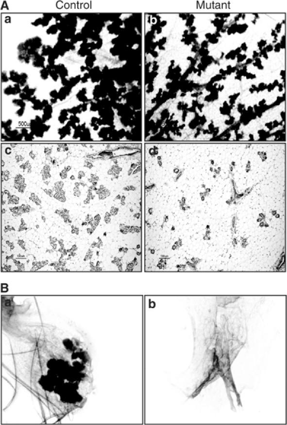
Integrin-extracellular matrix interactions play important roles in the coordinated integration of external and internal cues that are essential for proper development. To study the role of beta1 integrin in the mammary gland, Itgbeta1(flox/flox) mice were crossed with WAPiCre transgenic mice, which led to specific ablation of beta1 integrin in luminal alveolar epithelial cells. In the beta1 integrin mutant mammary gland, individual alveoli were disorganized resulting from alterations in cell-basement membrane associations. Activity of focal adhesion kinase (FAK) was also decreased in mutant mammary glands. Luminal cell proliferation was strongly inhibited in beta1 integrin mutant glands, which correlated with a specific increase of p21 Cip1 expression. In a p21 Cip1 null background, there was a partial rescue of BrdU incorporation, providing in vivo evidence linking p21 Cip1 to the proliferative defect observed in beta1 integrin mutant glands. A connection between p21 Cip1 and beta1 integrin as well as FAK was also established in primary mammary cells. These results point to the essential role of beta1 integrin signaling in mammary epithelial cell proliferation.


Figures

Figure 1
Figure 1
Specific ablation of β1 integrin in lactating mammary luminal epithelial cells. (A) Schematic representation of the floxed β1 integrin allele (a) and the recombined allele (b). The PCR primers for detection of the floxed and recombined allele and the product size are indicated. (c) The structure of the WAPiCre transgene is shown. (B–D) The 3-day lactating mammary glands of control or β1-integrin mutant mice were used for the following analyses. (B) Cre-mediated recombination was analyzed by PCR using genomic DNA. (C) β1 integrin and α-tubulin expression levels were analyzed by Western blotting on tissue lysates. (D) (a and b) Paraffin sections from (a) control mice or (b) β1 integrin mutant mice were analyzed for Cre recombinase expression using a specific antiserum. The black arrow in (b) indicates Cre positive cells. (c and d) Frozen sections from (c) control or (d) β1 integrin mutant mice were stained for β1 integrin. The white and yellow arrows indicate β1 integrin staining of luminal and myoepithelial cells, respectively. (E) Primary mammary cells were prepared from lactating control and β1 integrin mutant mice. The adhesion assay was performed on poly-D-Lysine (PDL), collagen type I (CI), fibronectin (FN), laminin (LN) and gelatin. After washing, the adhered cells were fixed, stained, solubilized and absorbance at 595 nm was read. The error bars represent standard deviation of the mean of triplicate samples within one experiment.
Figure 2
Figure 2
Impairment of alveolar integrity in β1 integrin mutant mammary glands. (A) Mammary glands were taken from control- and β1 integrin mutant mice during the first (a–f) or second (g and h) round of pregnancy and lactation, and paraffin sections were prepared and stained with H&E. The mammary glands were from mice at: day 16 of pregnancy (a and b); lactation day 1 (c and d) and day 5 (e–h). The black arrows in f and h indicate irregularities in the alveolar architecture; the yellow arrows in f and h indicate cells shed into the lumen of β1 integrin mutant mice. Shed cells, including those in the process thereof and those in the lumen, were quantified from three pairs of control and β1 integrin mutant mice at lactation day 5 by counting 1000 cells per section. Data (white boxes in e and f) are the mean±s.d. (B) Frozen sections of mammary glands from 5-day-lactating control mice (a, c, e and g) and β1 integrin mutant mice (b, d, f and h) mice were stained for: (a and b) α-smooth-muscle actin (× 20); (c and d) Npt2b (× 20); (e and f) Laminin-1 (× 20); (g and h) E-cadherin (× 60). The arrows in panels f and h indicate cells detached from the BM.
Figure 3
Figure 3
Loss of β1 integrin is not sufficient to increase apoptosis in luminal mammary epithelial cells. (A) Sections prepared from β1 integrin mutant mammary glands of 14-day lactating females were analyzed by electron microscopy. A detached cell in the lumen is outlined in white; another cell (black arrow) is in the process of detaching. Neither nucleus (N) is apoptotic (× 2500). (B) Paraffin sections from day-1 involuting mammary glands from control (a and c) and β1 integrin mutant (b and d) mice were H&E stained (a and b) (× 20) or TUNEL-stained (c and d) (× 20). Cells shed into the lumen (black arrows panel a and b) and TUNEL-positive cells (black arrows c and d) were quantified (insets on right), in sections from three pairs of control and β1 integrin mutant mice. In all cases, 900–1200 nuclei per section were counted and data are means±s.d. In (d) the green arrows show shed cell that are TUNEL-negative.
Figure 4
Figure 4
Loss of β1 integrin leads to reduced mammary cell proliferation, correlating with disrupted focal adhesion signaling and p21Cip1 upreguation. (A) Paraffin sections of mammary glands from 2-day lactating control and β1 integrin mutant mice were stained with a BrdU antibody and positive nuclei were quantified from three pairs of control and β1 integrin mutant females. In each case, 900–1200 nuclei per section were counted. Data are the mean±s.d. (B, C and E) Analysis of proteins extracts from 3- and 14-day lactating mammary glands. (B) Western blotting analysis revealed the levels of cyclins B1, D1 and E and CDK2. (C) Western blotting analysis revealed the levels of p21Cip1, p27Kip1 and p57Cip2; keratin 18 was used to control loading. (E) Western blotting analysis revealed the levels of Tyr397 FAK (PY397), FAK, vinculin, Tyr118 Paxillin (PY118) and paxillin. (D and F) Primary mammary cells were prepared from pregnant β1flox/flox mice (D) or FAKflox/flox mice (F) and infected with Adeno-β gal control virus or with Adeno-Cre-virus. Cell lysates were prepared and analyzed by Western blotting. The membranes were probed with antiserum specific for: (D) β1 integrin, Cre, p21Cip1, and p27Kip; Erk2 was used to control loading; (E) PY-397 FAK, FAK, p21Cip1 and Cre; actin was used to control loading.
Figure 5
Figure 5
Absence of p21Cip1 rescues the proliferation defect in β1 integrin mutant mice. Paraffin sections of mammary glands from 2-day lactating control, β1 integrin mutant and double p21Cip1null; β1 integrin mutant mice were stained with a BrdU antibody (B) and positive nuclei were quantified (A). Sections from three control, three β1 integrin mutant and seven double p21Cip1null; β1 integrin mutant females were examined. In each case, 900–1200 nuclei per section were counted and data are the mean±s.d. The asterik denotes significant changes (P⩽0.05) compared with β1 integrin mutant glands.
Figure 6
Figure 6
In MDA-MB-231 cells, loss of β1 integrin upregulates p21Cip1 expression and impairs proliferation. β1 integrin- or control lacZ-siRNA was transfected into MDA-MB-231 cells, 4 days later: (A) Western blotting for β1 integrin, p21Cip1 and α-tubulin was performed on cell lysates; (B) RNA was collected and p21Cip1 and actin mRNA levels were measured by quantitative PCR. (C) At 2 days after transfection of siRNA for LacZ or β1 integrin, MDA-MB-231 cultures were transfected with the WWP-Luc reporter plasmid plus control renilla plasmid and 2 days later p21Cip1 promoter activity was measured. Luciferase activity was normalized to the Renilla internal control and data are presented as the mean±s.d. (D and F) At 4 days after transfection of siRNA for LacZ, β1 integrin, p21Cip1+LacZ, or β1 integrin+p21Cip1 MDA-MB231 cell lysates were analyzed by Western blotting for p21Cip1, β1 integrin, and α-tubulin (F) or cells were counted in triplicate (D). Data are presented as the mean±s.d. (E) MDA-MB231 cells were transfected with LacZ siRNA and/or β1 integrin siRNA for 3 days, then labeled with BrdU for 1 h before collecting. Harvested cells were stained with anti-BrdU-FITC to quantitate cells in S phase and with 7AAD to stain DNA before flow cytometry was performed. The percent cells in G1 and G2–M is indicated in the top of each panel; the percentage of cells that incorporated BrdU is indicated in the box.
Figure 7
Figure 7
Wholemount and histological analysis of β1 integrin mutant mammary glands and mammary transplants. (A) Wholemount analysis (a–b) or histological analysis (c–d) of mammary glands taken from control and β1 integrin mutant females at day 16.5 of a second pregnancy. (Magnifications: a and b— × 6; c and d— × 10). (B) Wholemount analysis of mammary transplant outgrowths. Pieces of mammary tissue taken from 3-day lactating control mice (a) or β1-integrin mutant mice (b) were transplanted into cleared fat pads of 21-day-old syngeneic females. After 10 weeks the recipient female mice were mated and at pregnancy day-16 glands were removed and examined by wholemount analysis (× 8).

Similar articles

Cited by

References

    1. Aszodi A, Hunziker EB, Brakebusch C, Fassler R (2003) Beta1 integrins regulate chondrocyte rotation, G1 progression, and cytokinesis. Genes Dev 17: 2465–2479 - PMC - PubMed
    1. Barcellos-Hoff MH, Aggeler J, Ram TG, Bissell MJ (1989) Functional differentiation and alveolar morphogenesis of primary mammary cultures on reconstituted basement membrane. Development 105: 223–235 - PMC - PubMed
    1. Bill HM, Knudsen B, Moores SL, Muthuswamy SK, Rao VR, Brugge JS, Miranti CK (2004) Epidermal growth factor receptor-dependent regulation of integrin-mediated signaling and cell cycle entry in epithelial cells. Mol Cell Biol 24: 8586–8599 - PMC - PubMed
    1. Boudreau N, Sympson CJ, Werb Z, Bissell MJ (1995) Suppression of ICE and apoptosis in mammary epithelial cells by extracellular matrix. Science 267: 891–893 - PMC - PubMed
    1. Brakebusch C, Grose R, Quondamatteo F, Ramirez A, Jorcano JL, Pirro A, Svensson M, Herken R, Sasaki T, Timpl R, Werner S, Fassler R (2000) Skin and hair follicle integrity is crucially dependent on beta 1 integrin expression on keratinocytes. EMBO J 19: 3990–4003 - PMC - PubMed

Publication types

MeSH terms

LinkOut - more resources