. 2005 Jun 1;24(11):1931-41.
doi: 10.1038/sj.emboj.7600672. Epub 2005 May 5.

The yeast lipin Smp2 couples phospholipid biosynthesis to nuclear membrane growth

Affiliations

The yeast lipin Smp2 couples phospholipid biosynthesis to nuclear membrane growth

Helena Santos-Rosa et al. EMBO J. .

Abstract

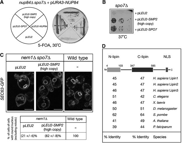
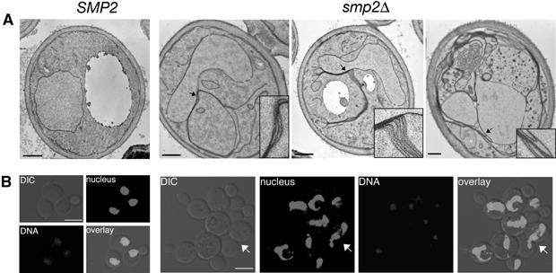
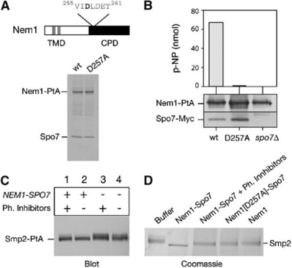
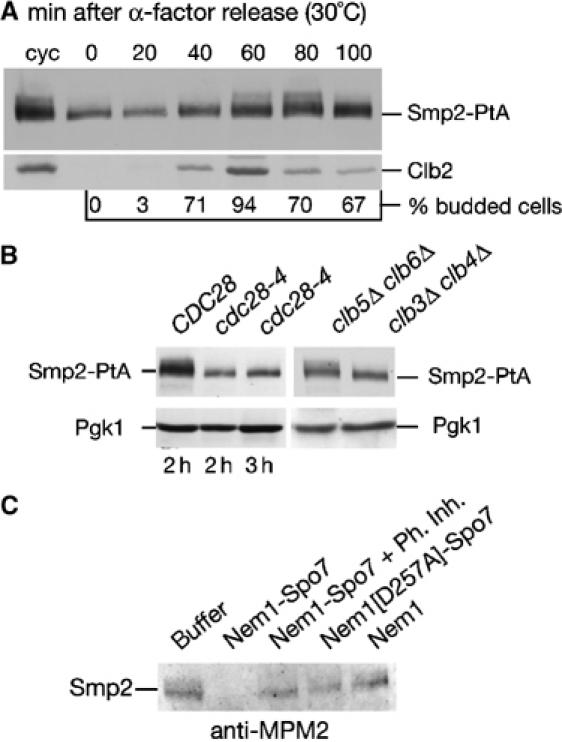
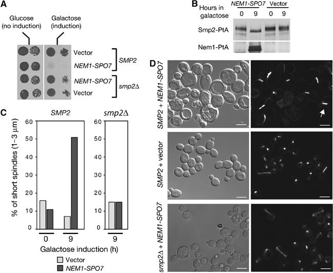
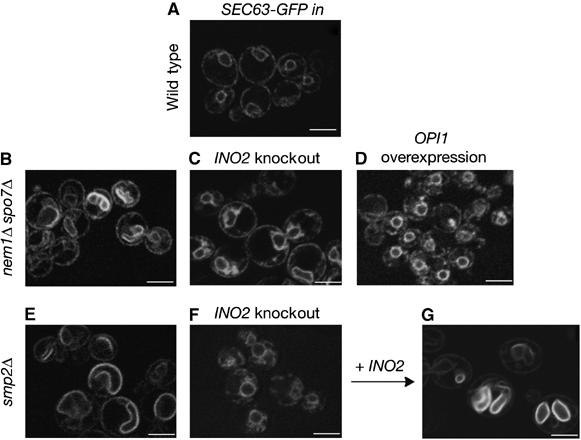
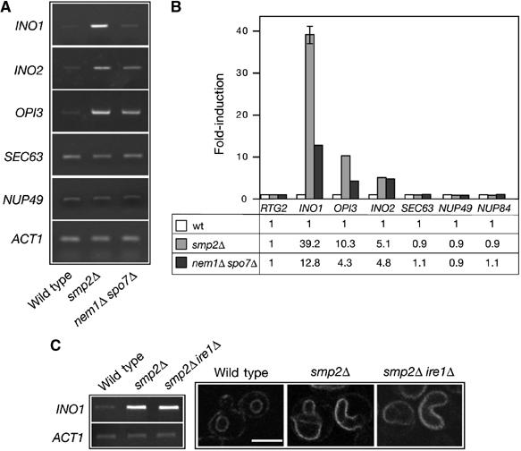
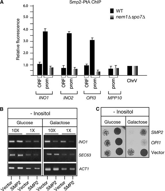
Remodelling of the nuclear membrane is essential for the dynamic changes of nuclear architecture at different stages of the cell cycle and during cell differentiation. The molecular mechanism underlying the regulation of nuclear membrane biogenesis is not known. Here we show that Smp2, the yeast homologue of mammalian lipin, is a key regulator of nuclear membrane growth during the cell cycle. Smp2 is phosphorylated by Cdc28/Cdk1 and dephosphorylated by a nuclear/endoplasmic reticulum (ER) membrane-localized CPD phosphatase complex consisting of Nem1 and Spo7. Loss of either SMP2 or its dephosphorylated form causes transcriptional upregulation of key enzymes involved in lipid biosynthesis concurrent with a massive expansion of the nucleus. Conversely, constitutive dephosphorylation of Smp2 inhibits cell division. We show that Smp2 associates with the promoters of phospholipid biosynthetic enzymes in a Nem1-Spo7-dependent manner. Our data suggest that Smp2 is a critical factor in coordinating phospholipid biosynthesis at the nuclear/ER membrane with nuclear growth during the cell cycle.


Figures

Figure 1
Figure 1
Functional interaction between NEM1–SPO7 and SMP2. (A) Identification of SMP2 as a high-copy number suppressor of the nup84Δ spo7Δ synthetic lethal mutant. The nup84Δ spo7Δ double deletion strain carrying a centromeric vector expressing NUP84 was transformed with the indicated plasmids. Transformants were grown on plates containing 5-FOA for 3 days. (B) SMP2 rescues the temperature-sensitive growth defect of spo7Δ cells. Spo7Δ cells transformed with the indicated plasmids were diluted in YEPD, spotted onto selective (-Leu) plates and grown at 37°C for 2 days. (C) Overexpression of SMP2 suppresses the nuclear membrane proliferation of nem1Δ spo7Δ cells. Upper panel: the nem1Δ spo7Δ mutant or the isogenic wild-type strain, expressing the ER marker Sec63-GFP were transformed with the indicated plasmids. Transformants were visualized by confocal microscopy. Bars, 5 μm. Lower panel: percentage of cells with no or small buds containing a round nucleus in wild-type and nem1Δ strains expressing GFP-Pus1. Three different transformants per strain were analyzed and for each one the number of cells counted was n=200. (D) Smp2 is evolutionarily conserved. Schematic representation of the primary structure of Smp2. The gray and black boxes indicate the highly conserved amino-terminal (N-lipin) and C-terminal (C-lipin) domains within Smp2. The percent sequence identity between the yeast domains and putative orthologues in various species is given.
Figure 2
Figure 2
Deletion of SMP2 induces nuclear membrane proliferation and nuclear expansion. (A) Thin section electron microscopy of wild-type (SMP2) or smp2Δ cells grown at 30°C and stained with potassium permanganate. Detail panels show enlargements of areas of the nuclear envelope in smp2Δ cells (highlighted by arrows) that display membrane proliferation. Bars, 0.5 μm. (B) DNA staining of wild-type (SMP2) or smp2Δ knockout cells expressing an intranuclear GFP-reporter (GFP-Pus1) used to depict nuclear structure (‘nucleus'). Cells were grown in selective medium at 30°C, fixed for 30 min and inspected by confocal microscopy. The white arrow points to a dividing yeast cell. Bars, 5 μm.
Figure 3
Figure 3
Dephosphorylation of Smp2 by the Nem1–Spo7 complex. (A) Upper panel: domain organization of Nem1. TMD, transmembrane domain. CPD, CTD Phosphatase domain. The DLD phosphoacceptor site is indicated. Lower panel: affinity purification of Nem1-PtA (‘wt') and Nem1[D257A]-PtA (‘D257A'). Purified proteins were analyzed by SDS–PAGE and Coomassie staining. The positions of the PtA-fusions and copurifying Spo7 are indicated. (B) The Nem1–Spo7 complex exhibits phosphatase activity in vitro. In vitro dephosphorylation of p-nitrophenylphosphate (p-NPP) by the Nem1–Spo7 complex. IgG-Sepharose beads loaded with (Nem1-PtA)–(Spo7-Myc), (Nem1[D257A]-PtA)–(Spo7-Myc) or Nem1-PtA fusions, were tested for the ability to hydrolyze p-NPP as described under Materials and methods. Absorbance of the generated p-nitrophenol (p-NP) was measured at 410 nm. The amount of Nem1 and Spo7 in each reaction was followed by Western blot with anti-PtA and anti-Myc antibodies respectively. (C) Nem1–Spo7 is a phosphatase for Smp2. Protein extracts from smp2Δ (lanes 1 and 2) or nem1Δ spo7Δ smp2Δ (lanes 3 and 4) strains expressing a Smp2-PtA fusion were prepared in the presence (lanes 1 and 3) or absence (lanes 2 and 4) of phosphatase inhibitors. Smp2-PtA was detected by western blot using anti-PtA antibody. (D) In vitro dephosphorylation of Smp2 by the Nem1–Spo7 complex. Native Smp2 (2 μg) was incubated with IgG-Sepharose beads alone (‘buffer') or beads containing the indicated protein A fusions for 30 min at 30°C. Reactions were resolved by 7% SDS–PAGE and Coomassie stained.
Figure 4
Figure 4
Smp2 is phosphorylated in a cell-cycle-dependent manner by Cdc28. (A) Phosphorylation of Smp2 takes place during mitosis. Cells expressing Smp2-PtA were synchronized by alpha-factor arrest/release. Protein extracts were prepared every 20 min and analyzed by Western blot using the indicated antibodies. Cell cycle stages were monitored by Clb2 cyclin levels and budding index. (B) Left panel: protein extracts from wild-type (‘CDC28') or cdc28-4 cells expressing Smp2-PtA grown for the indicated times at 37°C were analyzed by Western blot using anti-PtA antibodies. Right panel: protein extracts from clb3Δclb4Δ and clb5Δclb6Δ cells expressing Smp2-PtA were analyzed as above. (C) Native Smp2 is recognized by the MPM2 antibody in a Nem1–Spo7-dependent manner. Samples from the experiment in Figure 3D were transferred onto nitrocellulose membrane and incubated with the MPM2 antibody.
Figure 5
Figure 5
Accumulation of dephosphorylated Smp2 inhibits cell division. (A) Overexpression of the Nem1–Spo7 complex is lethal only in the presence of Smp2. Wild-type (‘SMP2') or smp2Δ cells transformed with centromeric vectors expressing NEM1 and SPO7 under the control of the GAL1/10 promoter, or with the corresponding empty vectors, were spotted onto selective plates supplemented with glucose or galactose and grown at 30°C. (B) Cells overexpressing NEM1SPO7 for the indicated times were analyzed for Smp2-PtA mobility shifts by Western blot using anti-PtA antibodies. (C) Wild-type or smp2Δ cells overexpressing NEM1SPO7 or empty vectors were scored for short spindles (1–3 μm) at the indicated times by using a tubulin-GFP reporter. (D) Cell (left panels) and spindle (right panels) morphologies using a tubulin-GFP fusion of wild-type or smp2Δ strains overexpressing NEM1SPO7 or empty vector for 21 h. The white arrow points to a typical short spindle. Bars, 5 μm.
Figure 6
Figure 6
Inhibition of the phospholipid biosynthetic pathway restores normal nuclear membrane structure in smp2Δ and nem1Δ spo7Δ cells. SEC63-GFP was used to visualize nuclear membrane structure in wild type (A), nem1Δ spo7Δ (B), nem1Δ spo7Δ ino2Δ (C), nem1Δ spo7Δ overexpressing OPI1 (D), smp2Δ (E), smp2Δ ino2Δ (F) and smp2Δ ino2Δ complemented by a plasmid expressing INO2 (G) strains. Transformants in early logarithmic phase were visualized by confocal microscopy. Bars, 5 μm.
Figure 7
Figure 7
Smp2 regulates expression of phospholipid biosynthetic genes. (A) Transcription of key genes involved in phospholipid biosynthesis is upregulated in smp2Δ and nem1Δ spo7Δ mutants. The mRNA levels of INO1, INO2, OPI3, SEC63, NUP49 and ACT1 were analyzed in the smp2Δ, nem1Δ spo7Δ and isogenic wild-type strains by semiquantitative RT–PCR. (B) As in (A), but the mRNA levels of INO1, INO2, OPI3, SEC63, NUP49 and NUP84 were analyzed by quantitative RT–PCR. Amplification of each sample was performed in triplicate and normalized to a control gene, RTG2, which is expressed at similar level to those analyzed and is unaffected by smp2Δ or nem1Δ spo7Δ mutations. Errors were less than 5% except for the smp2Δ INO1 sample. The fold-difference for the three strains is given below. (C) Upregulation of phospholipid biosynthesis in smp2Δ is independent of the unfolded protein response (UPR) pathway. The mRNA levels of INO1 and ACT1 were analyzed in the smp2Δ, smp2Δ ire1Δ and isogenic wild-type strains by RT–PCR (left panel). The nuclear morphology of smp2Δ, smp2Δ ire1Δ and isogenic wild-type cells expressing Sec63-GFP reporter was visualized by confocal microscopy (right panel). Bar, 5 μm.
Figure 8
Figure 8
Recruitment of Smp2 on the promoters of phospholipid biosynthetic genes. (A) Smp2 associates with the promoters of lipid biosynthetic genes. Chromatin inmunoprecipitation of Smp2-PtA was performed in wild-type and isogenic nem1Δ spo7Δ strains. The chromatin associated Smp2 was quantified by real-time PCR. Histogram bars represent relative fluorescence units, calculated as described under Materials and methods. An intergenic region in chromosome V was used as background control and arbitrarily given the value 1. (B) Transcription of INO1 is downregulated in cells overexpressing SMP2. The mRNA levels of INO1, SEC63 and ACT1 were analyzed by RT–PCR in cells overexpressing SMP2. Two different dilutions of the mRNA are shown. (C) Overexpression of Smp2 inhibits growth in media lacking inositol. Wild-type cells transformed with plasmids expressing SMP2 or OPI1 under the control of the GAL1/10 promoter or the respective empty vector, were spotted onto selective plates lacking inositol and supplemented with either glucose or galactose as carbon source and grown at 25°C.

Similar articles

Cited by

References

    1. Basco RD, Segal MD, Reed SI (1995) Negative regulation of G1 and G2 by S-phase cyclins of Saccharomyces cerevisiae. Mol Cell Biol 15: 5030–5042 - PMC - PubMed
    1. Bastos R, Lin A, Enarson M, Burke B (1996) Targeting and function in mRNA export of nuclear pore complex protein Nup153. J Cell Biol 134: 1141–1156 - PMC - PubMed
    1. Baumann O, Walz B (2001) Endoplasmic reticulum of animal cells and its organization into structural and functional domains. Int Rev Cytol 205: 149–214 - PubMed
    1. Burke B, Ellenberg J (2002) Remodelling the walls of the nucleus. Nat Rev Mol Cell Biol 3: 487–497 - PubMed
    1. Byers B, Goetsch L (1975) Behavior of spindles and spindle plaques in the cell cycle and conjugation of Saccharomyces cerevisiae. J Bacteriol 124: 511–523 - PMC - PubMed

Publication types

MeSH terms