. 2005 May 18;24(10):1787-97.
doi: 10.1038/sj.emboj.7600669. Epub 2005 May 5.

Urokinase-induced signaling in human vascular smooth muscle cells is mediated by PDGFR-beta

Affiliations

Urokinase-induced signaling in human vascular smooth muscle cells is mediated by PDGFR-beta

Julia Kiyan et al. EMBO J. .

Abstract

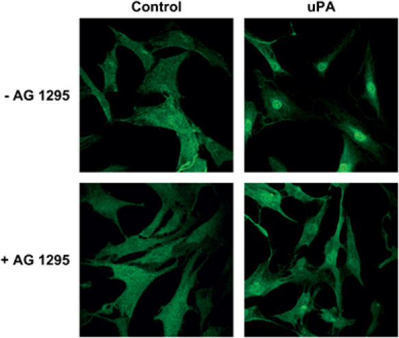
Urokinase (uPA)-induced signaling in human vascular smooth muscle cells (VSMC) elicits important cellular functional responses, such as cell migration and proliferation. However, how intracellular signaling is linked to glycolipid-anchored uPA receptor (uPAR) is unknown. We provide evidence that uPAR activation by uPA induces its association with platelet-derived growth factor receptor (PDGFR)-beta. The interaction results in PDGF-independent PDGFR-beta activation by phosphorylation of cytoplasmic tyrosine kinase domains and receptor dimerization. Association of the receptors as well as the tyrosine kinase activity of PDGFR-beta are decisive in mediating uPA-induced downstream signaling that regulates VSMC migration and proliferation. These findings provide a molecular basis for mechanisms VSMC use to induce uPAR- and PDGFR-directed signaling. The processes may be relevant to VSMC function and vascular remodeling.


Figures

Figure 1
Figure 1
Addition of uPA or ATF inhibits PDGF-BB-induced VSMC migration and proliferation. (A) VSMC migration stimulated by 25 nM uPA, 25 nM ATF, 20 ng/ml PDGF-BB: uPA+PDGF-BB or ATF+PDGF BB was assessed in a Boyden chamber (1 a.u. is 0.39 ODu). (B) VSMC proliferation stimulated as in (A) was assessed by BrdU uptake.
Figure 2
Figure 2
uPAR is required for uPA-dependent inhibition of PDGF-BB-induced migration and proliferation; PDGFR-β is required for uPA-induced responses. Expression of uPAR (A) and PDGFR-β (B) 24 h after cell nucleofection with corresponding silencing RNA duplexes and cells nucleofected with corresponding amount of nonspecific RNA duplexes assessed by Western blotting. The lower panels blotted with anti-actin antibodies demonstrate equal loading of the gels. Migration (C) and proliferation (D) of VSMC nucleofected with noncoding, uPAR and PDGFR-β si-RNA duplexes are shown. Cells were stimulated with 25 nM uPA, 20 ng/ml PDGF-BB. Migration was measured in a wounding model. Proliferation was measured by BrdU uptake.
Figure 3
Figure 3
PDGFR-β tyrosine kinase activity is required for uPA-induced responses. (A) VSMC migration was assessed in a Boyden chamber. Human VSMC were treated with indicated concentrations of AG1295 for 1 h and then allowed to migrate towards 25 nM uPA or 20 nM ATF (1 a.u. is 0.32 ODu). (B) VSMC proliferation was measured by BrdU uptake. Serum starved VSMC were stimulated as in (A) in the presence of indicated concentrations of AG1295. Migration (C) and proliferation (D) of VSMC expressing an R634 PDGFR-β mutant are shown. Migration was measured in a wounding model. Proliferation was measured by BrdU uptake. PDGFR-β was immunoprecipitated from VSMC stimulated with 10 ng/ml PDGF-BB (E) or 10 nM uPA (F), and the degree of its phosphorylation was assessed by Western blotting using anti-phosphotyrosine antibodies. The lower panels demonstrate equal amount of immunoprecipitated PDGFR-β.
Figure 4
Figure 4
uPAR and PDGFR-β in VSMC associate in an uPA-dependent manner. (A) PDGFR-β was immunoprecipitated from lysates of uPA-stimulated VSMC using rabbit polyclonal anti-PDGFR-β antibodies and uPAR in immunoprecipitates was visualized by Western blotting with R3 antibodies. (B) uPAR was immunoprecipitated from lysates of uPA-stimulated VSMC with R3 antibodies, and PDGFR-β in immunoprecipitates was visualized by Western blotting with rabbit polyclonal anti-PDGFR-β antibodies. (C) PDGFR-β was immunoprecipitated from lysates of PDGF-BB-stimulated VSMC, and uPAR in immunoprecipitates was visualized by Western blotting. (D) uPA-stimulated cells were subjected to crosslinking using 2.5 mM DSS, followed by immunoprecipitation with anti-PDGFR-β antibody. The immunoprecipitated proteins were then separated by PAGE and immunoblotted with anti-uPAR R3 antibody (left panel). Then the membranes were stripped and immunoblotted with anti- PDGFR-β antibodies (right panel). (E) VSMC were stimulated with uPA for 5 min in the presence of recombinant PDGFR/Fc chimera protein or nonspecific IgG, washed to remove unbound PDGFR/Fc, uPAR was immunoprecipitated and the amount of PDGFR-β in immunoprecipitates was analyzed by Western blotting.
Figure 5
Figure 5
uPAR and PDGFR-β in VSMC colocalize in an uPA-dependent manner. VSMC nonstimulated and stimulated with 10 nM uPA for the indicated time periods were immunostained with anti-PDGFR monoclonal antibodies and Alexa 488-conjugated secondary antibodies and anti-uPAR polyclonal antibodies visualized by Alexa 633-conjugated secondary antibodies. The frame size of all the images is 130 × 130 μm. The fluorograms were obtained by plotting each pixel of the overlay Alexa 488/Alexa 633 color fluorescence cell image. Pixel values of Alexa 488 and Alexa 633 fluorescence images represent horizontal and vertical axis values for each point, respectively. The points of the fluorograms are colored in accordance with count of pixels with specific Alexa 488 and Alexa 633 fluorescence intensities. The pixel count is shown in logarithmic scale.
Figure 6
Figure 6
uPA induces dimerization of PDGFR. VSMC were stimulated with uPA or PDGF-BB, then PDGFR was immunoprecipitated and the degree of receptor dimerization (A) and dimer phosphorylation (B) was assessed by Western blotting. (C) Serum-starved VSMC were stimulated for 5 min with uPA or PDGF-BB, then washed with cold PBS, fixed with 1% paraformaldehyde in PBS for 10 min at 4°C and labeled with mixture of Alexa 488- and Alexa 594-conjugated anti PDGFR-β antibodies, 5 μg/ml of each conjugate in PBS/1% BSA overnight at 4°C. The left panels show images of cells stained for PDGFR-β. The middle panels show the normalized FRET signal (FN) distributions over cells indicated by color coding. The right panels show histograms of the FN signal at the cell pixels.
Figure 7
Figure 7
PDGFR-β and its association with uPAR and PI3-K are required for uPA-induced RhoA activation and VSMC migration. (A) RhoA activation in response to uPA was measured by pulldown assay after VSMC pretreatment with 10 μM AG1295 or a corresponding amount of DMSO. (B) uPA-induced RhoA activation was measured in VSMC expressing either R634 PDGFR-β or F740/F751 PDGFR-β. (C) RhoA activation was measured in VSMC preincubated with 2 μg/ml PDGFR-β/Fc chimera protein prior to uPA stimulation. Migration (D) and proliferation (E) of VSMC expressing F740/F751 PDGFR-β mutant. Migration was measured in a wounding model. Proliferation was measured by BrdU uptake. Cells were stimulated by 25 nM uPA, 20 ng/ml PDGF-BB.
Figure 8
Figure 8
uPA-dependent STAT1 nuclear translocation requires active PDGFR-β. VSMC were pretreated with 10 μM AG1295 for 1 h, then stimulated with 10 nM uPA for 35 min and immunostained with anti-STAT1 polyclonal antibodies visualized byAlexa 488-conjugated secondary antibodies. The frame size of all the images is 240 × 240 μm.
Figure 9
Figure 9
uPA-induced STAT1 (Tyr701) phosphorylation requires active PDGFR-β. (A) Nontreated VSMC and cells pretreated with 10 μM AG1295 were stimulated with 10 nM uPA for indicated time. STAT1 (Tyr701) phosphorylation was assessed by Western blotting with anti-phospho-(Tyr701) STAT1 antibodies. The lower panels demonstrate equal protein loading of the gels. uPA-dependent STAT1 (Tyr701) phosphorylation in VSMC expressing R634 (B) and F740/751 (C) mutants, measured by Western blotting with anti-phospho-(Tyr701) STAT1 antibodies. The lower panels demonstrate equal protein loading of the gels. (D) STAT1 phosphorylation in response to uPA measured in VSMC preincubated with 2 μg/ml PDGFR-β/Fc chimera protein prior to uPA stimulation. The lower panel demonstrates equal protein loading of the gel.

References

    1. Banai S, Wolf Y, Golomb G, Pearle A, Waltenberger J, Fishbein I, Schneider A, Gazit A, Perez L, Huber R, Lazarovichi G, Rabinovich L, Levitzki A, Gertz DT (1998) PDGF-receptor tyrosine kinase blocker AG1295 selectively attenuates smooth muscle cell growth in vitro and reduces neointimal formation after balloon angioplasty in swine. Circulation 97: 1960–1969 - PubMed
    1. Blasi F, Carmeliet P (2002) uPAR: a versatile signalling orchestrator. Nat Rev Mol Cell Biol 3: 932–943 - PubMed
    1. Bromberg JF, Fan Z, Brown C, Mendelsohn J, Darnell JE Jr (1998) Epidermal growth factor-induced growth inhibition requires Stat1 activation. Cell Growth Differ 9: 505–512 - PubMed
    1. Carlin SM, Roth M, Black JL (2003) Urokinase potentiates PDGF-induced chemotaxis of human airway smooth muscle cells. Am J Physiol Lung Cell Mol Physiol 284: L1020–L1026 - PubMed
    1. Carmeliet P, Moons L, Herbert JM, Crawley J, Lupu F, Lijnen R, Collen D (1997) Urokinase but not tissue plasminogen activator mediates arterial neointima formation in mice. Circ Res 81: 829–839 - PubMed

Publication types

MeSH terms

Substances