. 2005 May 18;24(10):1798-809.
doi: 10.1038/sj.emboj.7600660. Epub 2005 May 5.

Prostaglandin D2 induces nuclear import of the sex-determining factor SOX9 via its cAMP-PKA phosphorylation

Affiliations

Prostaglandin D2 induces nuclear import of the sex-determining factor SOX9 via its cAMP-PKA phosphorylation

Safia Malki et al. EMBO J. .

Abstract

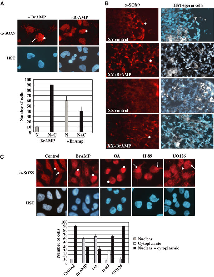
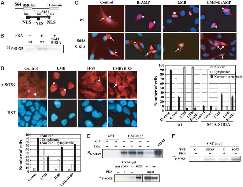
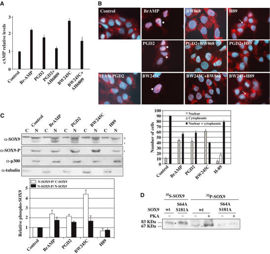
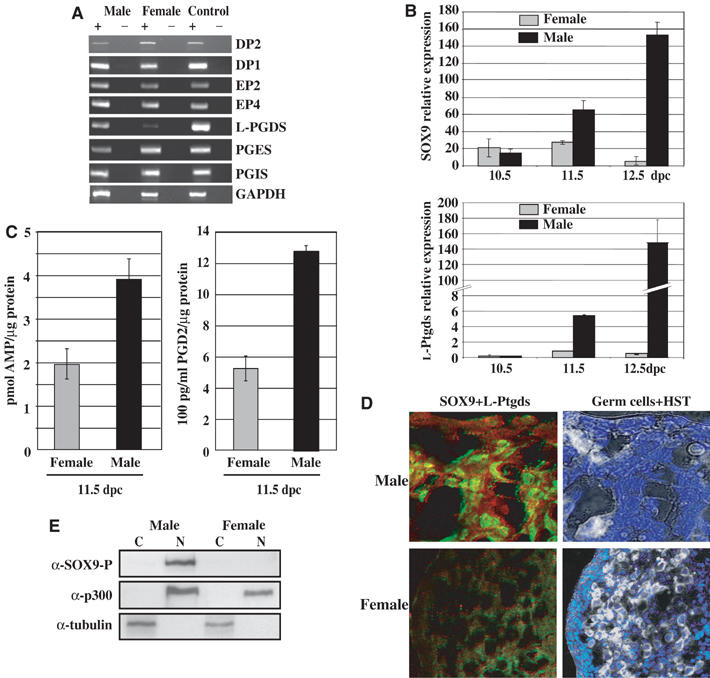
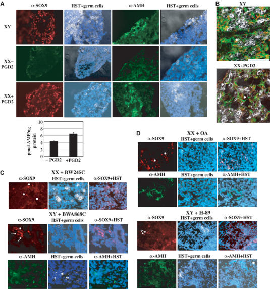
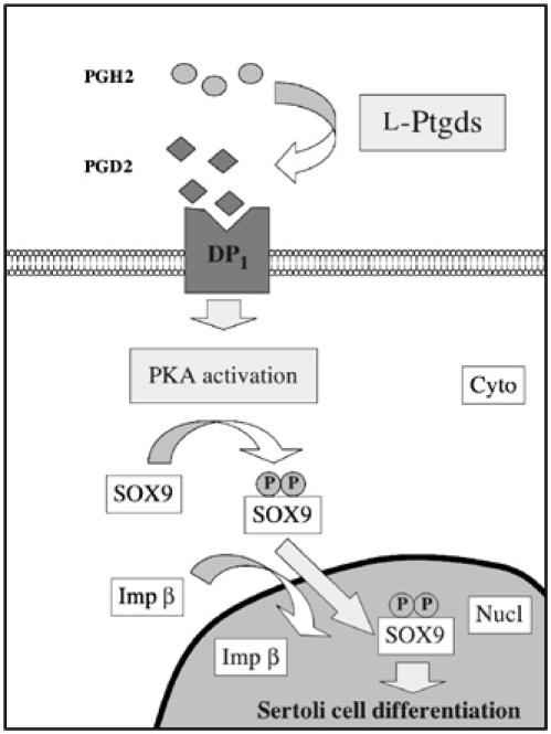
During mammalian gonadal development, nuclear import/export of the transcription factor SOX9 is a critical step of the Sry-initiated testis-determining cascade. In this study, we identify a molecular mechanism contributing to the SOX9 nuclear translocation in NT2/D1 cells, which is mediated by the prostaglandin D2 (PGD2) signalling pathway via stimulation of its adenylcyclase-coupled DP1 receptor. We find that activation of cAMP-dependent protein kinase A (PKA) induces phosphorylation of SOX9 on its two S64 and S181 PKA sites, and its nuclear localization by enhancing SOX9 binding to the nucleocytoplasmic transport protein importin beta. Moreover, in embryonic gonads, we detect a male-specific prostaglandin D synthase expression and an active PGD2 signal at the time and place of SOX9 expression. We thus propose a new step in the sex-determining cascade where PGD2 acts as an autocrine factor inducing SOX9 nuclear translocation and subsequent Sertoli cell differentiation.


Figures

Figure 1
Figure 1
SOX9 nuclear translocation is induced by stimulation of the cAMP–PKA pathway. (A) NT2/D1 cells express SOX9 in the nuclear (*) and the cytoplasmic (→) compartments as shown by immunostaining with an anti-SOX9 antibody (α-SOX9) and DNA staining (HST) (magnification × 60). Statistical SOX9 subcellular distribution was represented on the corresponding graph: nuclear SOX9 (N, gray bars) and nuclear+cytoplasmic SOX9 (N+C, black bars). (B) cAMP pathway stimulation in cultured mouse gonads induces SOX9 nuclear localization in female gonads. Dissected pairs of mesogonads from 10.5 dpc embryos were cultured with Br-AMP for 24 h. Nuclear SOX9 (in red, *) was visualized with a specific antibody, while germ cells (in white) and nuclei (in blue) were merged on the same sections (HST+germ cells) (magnification × 40). (C) PKA activation is involved in SOX9 nuclear translocation. Following treatment of NT2/D1 cells with different drugs, protein kinase inhibitors H-89 (PKA), UO126 (MAPK), phosphatase inhibitor OA and Br-AMP, SOX9 subcellular distribution was analysed by immunostaining with an anti-SOX9 antibody (α-SOX9) and DNA staining (HST) (magnification × 60). Nuclear SOX9 is indicated by an (*) and cytoplasmic SOX9, by an (→). Statistical SOX9 subcellular distribution was represented in the corresponding graph: nuclear SOX9 (grey bars), cytoplasmic SOX9 (white bars) and nuclear+cytoplasmic SOX9 (black bars).
Figure 2
Figure 2
cAMP–PKA stimulation induces SOX9 nuclear translocation via facilitating its nuclear import. (A) Position and spacing of PKA phosphorylation sites (S64 and S181) in relation to the nuclear localization (NLS) and nuclear export (NES) sequences within SOX9 full sequence. (B) Phosphorylation state of wt and S64A–S181A mutant SOX9 TNT proteins was controlled by in vitro PKA labelling (+) in the presence of γ32P-ATP after α-Flag immunoprecipitation and autoradiography. (C) Subcellular distribution of wt and mutant SOX9 protein in NIH3T3 cells. Transfected cells were treated with Br-AMP (BrAMP), LMB or both (LMB+BrAMP), and SOX9 revealed by α-SOX9 immunostaining (red) was co-localized with HST staining (blue) (magnification × 60). (D) PKA phosphorylation increases SOX9 nuclear import without affecting its nuclear export. NT2/D1 cells were treated with LMB, H-89 or LMB+H-89, and SOX9 subcellular distribution was analysed by immunofluorescence with α-SOX9 antibody (red) and DNA staining (HST, blue) (magnification × 60). In both panels (C, D), nuclear SOX9 is indicated by an (*) and cytoplasmic SOX9 is indicated by an (→), and statistical SOX9 subcellular distribution is represented in the corresponding graphs. (E) Effect of SOX9 phosphorylation on its interaction with importin β. GST pulldown experiments were performed using GST alone or GST-importin β proteins and in vitro translated 35S Met-wt or S64A–S181A (mut) SOX9. Prior to the binding reaction, SOX9 protein was subjected to incubation in the presence (+) or absence (−) of PKA or calf intestine phosphatase (CIP) (panels 35S-SOX9). Input represent 50% of the SOX9 TNT proteins. (F) The phosphorylation state of SOX9 required for its interaction with importin β was confirmed in GST pulldown experiment using 32P-labelled SOX9. Cold TNT-SOX9 (SOX9) or TNT-pcDNA (C) was submitted to in vitro phosphorylation by PKA (+) in the presence of γATP prior to the binding reaction. 32P panel shows autoradiography of importin β-bound SOX9 after GST pulldown reactions.
Figure 3
Figure 3
PGD2 pathway induces SOX9 nuclear translocation in NT2/D1 cells. (A) cAMP Elisa assays (R&D systems) were performed on NT2/D1 lyzates treated with Br-AMP, PGD2 and its agonist (BW245C) or antagonist (AH6809). Relative cAMP levels are expressed by nmol of produced cAMP per ng of total proteins and are normalized to the basal response of untreated cells (control). (B) SOX9 subcellular distribution in NT2/D1 cells was analysed after treatment with Br-AMP, PGD2 and its DP1-agonist (BW245C) and DP1-specific antagonist (BWA868C) or PKA inhibitor H-89. SOX9 localization was detected by α-SOX9 immunostaining (red) merged with nuclei staining (blue). Nuclear SOX9 is denoted by an (*) and cytoplasmic SOX9 by an (→) and statistical SOX9 subcellular distribution was represented in the corresponding graph. (C) SOX9 phosphorylation status was detected by Western blotting with an anti-phospho SOX9 antibody (α-SOX9-P) on sub-cellular fractions (cytoplasmic, C; nuclear, N) of treated NT2/D1 cells while α-SOX9 blot determined whole SOX9 expression. (*) and (°) indicate non-phosphorylated and phosphorylated SOX9, respectively. Relative levels of phospho-SOX9 protein induced by prostanoid treatments were quantified by the NIH Image program. Cytoplasmic or nuclear SOX9 were normalized to α-tubulin and α-p300, respectively. The ratios nuclear phospho-SOX9 (N-SOX9-P)/cytoplasmic SOX9 (C-SOX9) (white bars) and nuclear phospho-SOX9 (N-SOX9-P)/nuclear non-phospho-SOX9 (N-SOX9) (black bars) were represented on the adjacent histogram. (D) Determination of the phosphorylation state of SOX9 isoforms. Cold wt SOX9 and S64A–S181A SOX9 TNT products were subjected (+) or not (−) to in vitro PKA phosphorylation in presence of γ32P-ATP (32P panel). Migration of phosphorylated (band °) SOX9 proteins were compared to that of nonphosphorylated (band *) SOX9 proteins (35S panel).
Figure 4
Figure 4
PGD2 pathway is expressed in mouse embryonic gonads at the time of sexual differentiation. (A) Prostanoid receptor and prostaglandin synthase expression was evaluated by semiquantitative RT–PCR on total RNAs isolated from 11.5 dpc embryonic gonads in the presence (+) or absence (−) of SuperScriptII and using the primers indicated in Table I. Control experiments were performed on total RNA from whole 11.5 dpc male and female mouse embryos. (B) L-Ptgds and SOX9 relative expressions were compared in SF-1-positive cells from 10.5, 11.5 and 12.5 dpc male and female gonads by quantitative RT–PCR using the primers indicated in Table I. They were measured in fluorescence units and normalized to Hprt expression, in triplicates on two independent RNA preparations. (C) Intracellular cAMP and PGD2 levels were measured in differentiating 11.5 dpc mouse gonads using the cAMP Elisa kit (R&D systems) and the Prostaglandin D2-MOX Express EIA kit (Cayman Chemical), respectively. cAMP and PGD2 levels were measured in triplicates in two experiments, relatively to protein concentration, and are represented by black bars (male) and grey bars (female). (D) L-Ptgds protein was detected in 11.5 dpc mouse male and female gonad sections by immunofluorescence with an anti-L-Ptgds antibody (1/100). Cytoplasmic L-Ptgds was detected (red) in SOX9-expressing cells (green) as visualized by confocal microscopy (magnification × 100 for male and × 40 for female). Germ cells (in white) were merged with HST DNA staining (blue). (E) Localization of phosphorylated SOX9 in subcellular fractions of 13.5 dpc male and female gonads. After fractionation, phospho-SOX9 was detected in nuclear fraction (N) using an anti-SOX9-P antibody. Western blotting with α-tubulin and α-p300 antibodies identified cytoplasmic (C) and nuclear (N) fractions, respectively.
Figure 5
Figure 5
PGD2 via its DP1 receptor induces SOX9 nuclear localization and AMH expression in cultured female gonads. Dissected pairs of gonads from 8 to 15 TS (10.5–11.5 dpc) male (XY) and female (XX) embryos were cultured ex vivo (A, B) with (+PGD2) or without PGD2 (−PGD2) for 3 days, (C) with BW245C or BWA868C, (D) with OA or H-89 for 3 days. Nuclear and cytoplasmic SOX9 (red) and AMH (green) were revealed by specific antibodies and visualized by fluorescence microscopy (A, magnification × 40, C, D, magnification × 60). cAMP stimulation in PGD2-treated (8 to 15 TS) XX gonads was measured relatively to the protein concentration of the extracts and represented on the corresponding graph. (B) Expression of AMH in SOX9-positive cells was recorded on a confocal microscope after double immunostaining (Adams and McLaren, 2002) of male (XY) and treated female gonads (XX+PGD2) (scale bar=100 μm). Germ cells (white) were merged with DNA staining (HST, blue) when indicated.
Figure 6
Figure 6
Model for the regulation of SOX9 nuclear localization through activation of PGD2/PKA pathway. Cyto and Nucl indicate cytoplasmic and nuclear compartments, respectively. P represents phosphorylated serines (Ser64 and Ser181) on SOX9 protein.

References

    1. Adams IR, McLaren A (2002) Sexually dimorphic development of mouse primordial germ cells: switching from oogenesis to spermatogenesis. Development 129: 1155–1164 - PubMed
    1. Arango NA, Lovell-Badge R, Behringer RR (1999) Targeted mutagenesis of the endogenous mouse Mis gene promoter: in vivo definition of genetic pathways of vertebrate sexual development. Cell 99: 409–419 - PubMed
    1. Argentaro A, Sim H, Kelly S, Preiss S, Clayton A, Jans DA, Harley VR (2003) A SOX9 defect of calmodulin-dependent nuclear import in campomelic dysplasia/autosomal sex reversal. J Biol Chem 278: 33839–33847 - PubMed
    1. Boie Y, Sawyer N, Slipetz DM, Metters KM, Abramovitz M (1995) Molecular cloning and characterization of the human prostanoid DP receptor. J Biol Chem 270: 18910–18916 - PubMed
    1. Brennan J, Tilmann C, Capel B (2003) Pdgfr-alpha mediates testis cord organization and fetal Leydig cell development in the XY gonad. Genes Dev 17: 800–810 - PMC - PubMed

Publication types

MeSH terms