. 2005 May 18;24(10):1775-86.
doi: 10.1038/sj.emboj.7600658. Epub 2005 May 5.

Sec17p and HOPS, in distinct SNARE complexes, mediate SNARE complex disruption or assembly for fusion

Affiliations

Sec17p and HOPS, in distinct SNARE complexes, mediate SNARE complex disruption or assembly for fusion

Kevin M Collins et al. EMBO J. .

Abstract

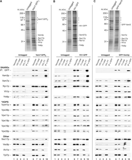
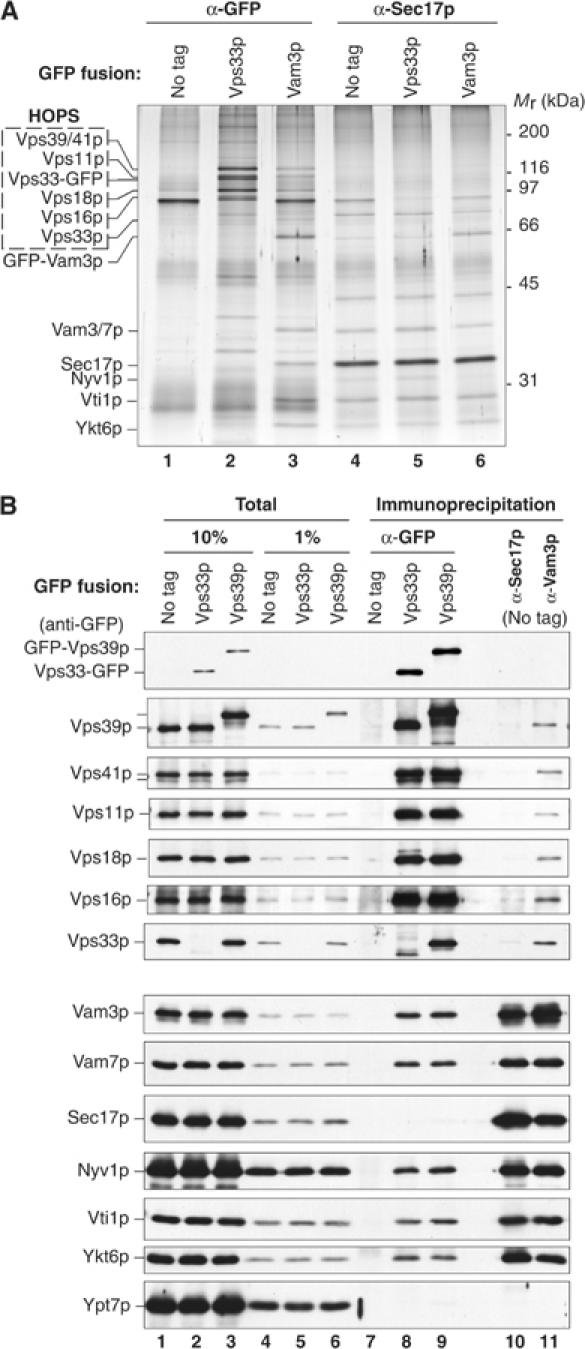
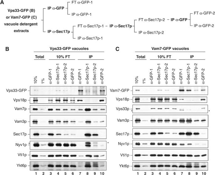
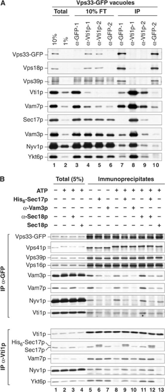
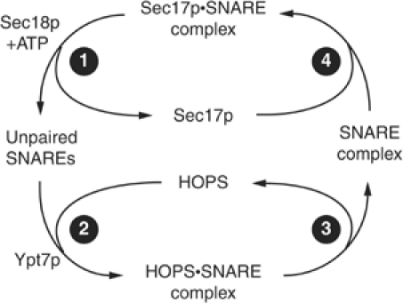
SNARE functions during membrane docking and fusion are regulated by Sec1/Munc18 (SM) chaperones and Rab/Ypt GTPase effectors. These functions for yeast vacuole fusion are combined in the six-subunit HOPS complex. HOPS facilitates Ypt7p nucleotide exchange, is a Ypt7p effector, and contains an SM protein. We have dissected the associations and requirements for HOPS, Ypt7p, and Sec17/18p during SNARE complex assembly. Vacuole SNARE complexes bind either Sec17p or the HOPS complex, but not both. Sec17p and its co-chaperone Sec18p disassemble SNARE complexes. Ypt7p regulates the reassembly of unpaired SNAREs with each other and with HOPS, forming HOPS.SNARE complexes prior to fusion. After HOPS.SNARE assembly, lipid rearrangements are still required for vacuole content mixing. Thus, Sec17p and HOPS have mutually exclusive interactions with vacuole SNAREs to mediate disruption of SNARE complexes or their assembly for docking and fusion. Sec17p may displace HOPS from SNAREs to permit subsequent rounds of fusion.


Figures

Figure 1
Figure 1
Isolation of SNARE complexes from yeast vacuoles. Vacuoles isolated from (A) untagged (DKY6281) and Vam7-GFP2, (B) untagged (BJ2168) and Vti1-GFP, and (C) untagged (BJ3505) and GFP-Vam3p-tagged yeast strains were solubilized and immunoprecipitated with antibodies to GFP (see Materials and methods). Immunoprecipitates were analyzed by SDS–PAGE, silver staining (top), and immunoblotting (bottom). Vacuoles were prepared by large-scale fermentation and batch purification (Ungermann et al, 1999; Seals et al, 2000). Vacuoles in PS buffer (120 μg) were reisolated by centrifugation (11 000 g, 20 min, 4°C). Pellets were suspended in solubilization buffer (450 μl; see Materials and methods). In all, 10% of the suspension (vacuoles, lanes 1 and 6) and high-speed supernatants (10% total, lanes 2 and 7; 1% total, lanes 3 and 8) were removed prior to immunoprecipitation (overnight, 4°C) with immobilized antibodies to GFP. The beads were collected, and 10% of the flow-through supernatant was removed (10% FT, lanes 4 and 9). Collected beads were resuspended five times with solubilization buffer and bound material was eluted by heating in sample buffer (IP, lanes 5 and 10). The samples were separated by SDS–PAGE and silver stained (top) or transferred to nitrocellulose and decorated with antibodies (bottom). Polypeptides identified by immunoblot are indicated; HOPS subunits are denoted by asterisks.
Figure 2
Figure 2
Complexes of HOPS and multiple vacuole SNAREs lack Sec17p. (A) Vacuoles isolated from untagged (BJ2168), Vps33-GFP-tagged, and GFP-Vam3p-tagged yeast strains (300 μg each) were sedimented (11 000 g, 20 min, 4°C) and suspended in 1 ml of solubilization buffer (see Materials and methods). A portion of each extract and an equivalent volume of the high-speed supernatant were removed, and the remaining extracts were divided in half for immunoprecipitation (1 h, 4°C) with affinity-purified antibodies to GFP (lanes 1–3) or Sec17p (lanes 4–6). The immunoprecipitated material was separated by SDS–PAGE and silver stained. (B) Vps33p and Vps39p are similarly associated with vacuole SNAREs but not Sec17p. Vacuoles (120 μg) bearing no GFP fusion protein (DKY6281), or Vps33-GFP or GFP-Vps39p were solubilized (see Materials and methods) and immunoprecipitated with affinity-purified antibodies to GFP (lanes 7–9), Sec17p (lane 10), or Vam3p (lane 11).
Figure 3
Figure 3
Immunodepletion of Sec17p does not affect the proportion of SNAREs found associated with HOPS. (A) The Sec17p immunodepletion strategy. Detergent extracts of vacuoles bearing either Vps33-GFP (B) or Vam7-GFP (C) were prepared (Total, lanes 1 and 2) and immunoprecipitated with antibodies to GFP (IP α-GFP-1, lane 7) or Sec17p (IP α-Sec17p-1, lane 8). The immunodepleted extracts from the first anti-GFP and anti-Sec17p immunoprecipitations were designated flow-through (FT α-GFP-1, lane 3; FT α-Sec17p-1, lane 4). The FT α-Sec17p-1 extract was immunodepleted again with fresh antibodies to Sec17p (IP α-Sec17p-2, lane 9). The twice depleted material was decanted, and an aliquot saved as FT α-Sec17p-2 (lane 5). The extract was then immunoprecipitated with antibodies to GFP (IP α-GFP-2, lane 10), and an aliquot of the material remaining after this immunoprecipitation was removed (FT α-GFP-2, lane 6). After immunoprecipitations, bound material was eluted by heating in reducing sample buffer for SDS–PAGE and immunoblotting. Asterisks indicate inefficiently stripped immunoreactive material leftover from previous antibody incubations.
Figure 4
Figure 4
Sec17p can displace HOPS from interactions with SNARE complexes. (A) HOPS interacts with vacuole SNARE complexes. As outlined in Figure 3A, detergent extracts of vacuoles bearing Vps33-GFP (300 μg) were prepared and samples representing 10 and 1% of the extract (Total, lanes 1 and 2) were removed. The extracts were then divided. One-half was immunoprecipitated with antibodies to GFP (IP α-GFP-1, lane 7). The other extract was sequentially immunodepleted (1 h each) with immobilized antibodies to Vti1p (IP α-Vti1p-1, lane 8; IP α-Vti1p-2, lane 9) and then immunoprecipitated with antibodies to GFP (IP α-GFP-2, lane 10). After each step, samples were assayed for the extent of immunodepletion (see Figure 3A). After immunoprecipitation, bound material was eluted by boiling in sample buffer for SDS–PAGE and immunoblotting. (B) Sec17p displaces SNAREs from HOPS. Vacuoles (60 μg) bearing Vps33-GFP were incubated with buffer, anti-Sec18p Fab, anti-Vam3p Fab, or Sec18p (10 μg/ml) for 5 min on ice. Sec17p (24 μg/ml) was added where indicated and the reactions were further incubated for 5 min. Samples were normalized to fusion reaction salt conditions (all treatments) and an ATP regenerations system (see Materials and methods) was added where indicated. Reactions were incubated at 27°C (15 min), placed on ice (5 min), and vacuoles were reisolated (11 000 g, 10 min, 4°C). Pellets were extracted with solubilization buffer (400 μl), and insoluble material was removed (see Materials and methods). A sample of each solubilized extract was heated in sample buffer (Total, 5%). From one-half of the extract, Vps33-GFP was immunoprecipitated with antibodies to GFP and the other half was immunoprecipitated with antibodies to Vti1p (16 h, 4°C).
Figure 5
Figure 5
HOPS·SNARE complexes assemble during docking and prior to fusion. (A) Vps33p functions during docking. Standard vacuole fusion reactions were begun and, at indicated times, aliquots were dispensed to tubes on ice or to tubes with buffer or antibodies to Sec18p, Vps33p, or Vam3p. The reactions were further incubated at 27°C and assayed for alkaline phosphatase activity after 90 min. (B) Testing Ypt7p and HOPS requirements for fusion and SNARE complex assembly in the absence of Sec17/18p function. Fusion reactions (180 μl) contained anti-Sec17p antibodies and ATP. After 15 min, inhibitors (Gdi1p/Gyp1-46p, affinity-purified antibodies to Vps33p, Fab fragments of antibody to Vam3p, or MED) were added and incubated (5 min) before GST-Vam7p (190 ng) was added where indicated. Reactions were continued for a total of 70 min and then placed on ice. An aliquot (30 μl) was removed to assay matured alkaline phosphatase activity (C), and the associations of the added GST-Vam7p were assessed by precipitation from vacuole detergent extracts using immobilized glutathione (D). Vacuoles were sedimented from the remainder of the reactions (11 000 g, 10 min, 4°C) and the pellets were extracted on ice with solubilization buffer with 1 mM dithiothreitol (DTT) (200 μl; see Materials and methods). Detergent-insoluble material was removed by ultracentrifugation, and GST-Vam7p was retrieved with glutathione Sepharose (20 μl beads, 1 h, 4°C). Unbound material was removed as described for immunoprecipitations (see Materials and methods), and bound material was eluted by heating in reducing sample buffer.
Figure 6
Figure 6
Resolution of SNARE complex assembly and vacuole fusion. (A) Separation of SNARE and lipid ligand inhibitor sensitivity during fusion reactions driven by added Vam7p. Fusion reactions contained antibodies to Sec17p and ATP. Vam7p (6.3 μg/ml) was added after 20 min. Reactions received inhibitors (anti-Vam3p IgG, MED, ENTH, or placed on ice) at indicated times (19.5, 20.25, 20.75, 22, 25, 35, or 90 min), and alkaline phosphatase activity was measured after 90 min. PMSF (7 mM final concentration) was added to reactions placed on ice to terminate further proteolytic maturation of Pho8p. (B–E) Lipid ligands prevent vacuole fusion. As in (A), fusion reactions were begun at 27°C in the presence of anti-Sec17p antibody (B). The block was bypassed with Vam7p (C) after 20 min, and after 15 s, MED (D) or ENTH (E) was added. After 90 min, fusion was assayed by alkaline phosphatase activity (ALP) or fluorescence microscopy. Reactions (30 μl) were mixed with low-melt agarose (50 μl) containing the lipophilic dye MDY-64 and 10 μl of the suspension was mounted on a glass slide. Fluorescent micrographs were taken from random fields, and the surface areas of individual vacuoles were determined (see Materials and methods). A representative image from one field is shown (inset; scale bar is 5 μm).
Figure 7
Figure 7
Working model of the HOPS and Sec17p cycles.

Similar articles

Cited by

References

    1. Allan BB, Moyer BD, Balch WE (2000) Rab1 recruitment of p115 into a cis-SNARE complex: programming budding COPII vesicles for fusion. Science 289: 444–448 - PubMed
    1. Ausubel FM, Brent R, Kingston RE, Moore DD, Seidman JG, Smith JA, Struhl K (1994) Current Protocols in Molecular Biology. New York: John Wiley & Sons Inc.
    1. Boeddinghaus C, Merz AJ, Laage R, Ungermann C (2002) A cycle of Vam7p release from and PtdIns 3-P-dependent rebinding to the yeast vacuole is required for homotypic vacuole fusion. J Cell Biol 157: 79–89 - PMC - PubMed
    1. Bryant NJ, James DE (2003) The Sec1p/Munc18 (SM) protein, Vps45p, cycles on and off membranes during vesicle transport. J Cell Biol 161: 691–696 - PMC - PubMed
    1. Cao X, Ballew N, Barlowe C (1998) Initial docking of ER-derived vesicles requires Uso1p and Ypt1p but is independent of SNARE proteins. EMBO J 17: 2156–2165 - PMC - PubMed

Publication types

MeSH terms

Substances