Comparative Study
. 2005 Jun 1;24(11):1901-10.
doi: 10.1038/sj.emboj.7600657. Epub 2005 May 12.

H662 is the linchpin of ATP hydrolysis in the nucleotide-binding domain of the ABC transporter HlyB

Affiliations
Comparative Study

H662 is the linchpin of ATP hydrolysis in the nucleotide-binding domain of the ABC transporter HlyB

Jelena Zaitseva et al. EMBO J. .

Abstract

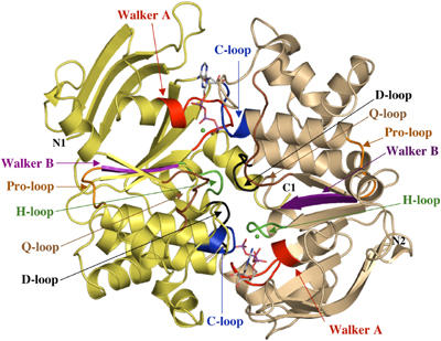
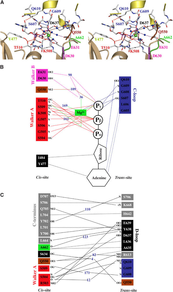
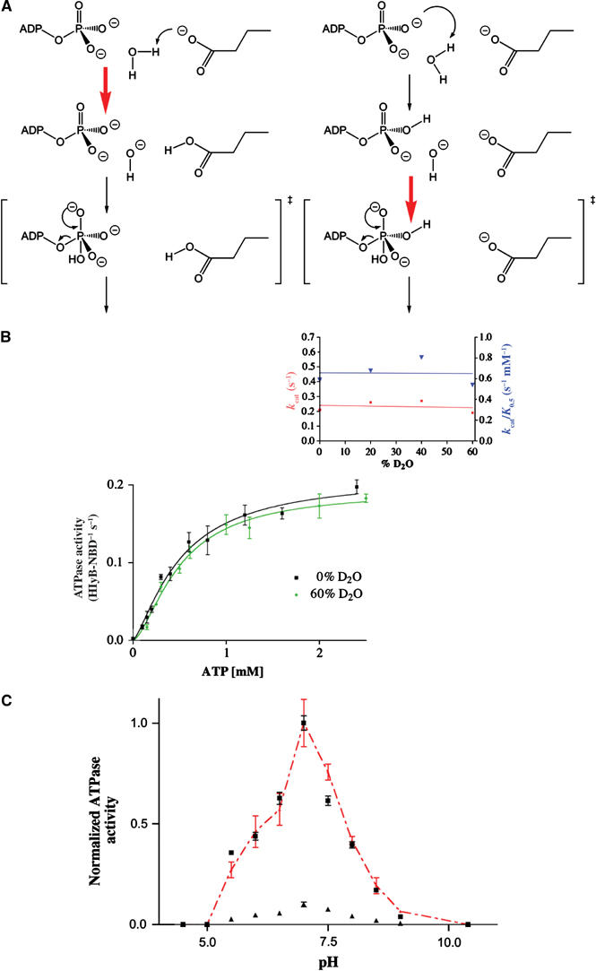
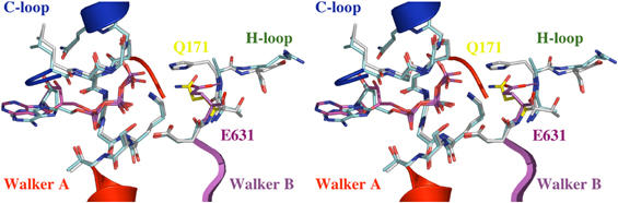
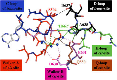
The ABC transporter HlyB is a central element of the HlyA secretion machinery, a paradigm of Type I secretion. Here, we describe the crystal structure of the HlyB-NBD (nucleotide-binding domain) with H662 replaced by Ala in complex with ATP/Mg2+. The dimer shows a composite architecture, in which two intact ATP molecules are bound at the interface of the Walker A motif and the C-loop, provided by the two monomers. ATPase measurements confirm that H662 is essential for activity. Based on these data, we propose a model in which E631 and H662, highly conserved among ABC transporters, form a catalytic dyad. Here, H662 acts as a 'linchpin', holding together all required parts of a complicated network of interactions between ATP, water molecules, Mg2+, and amino acids both in cis and trans, necessary for intermonomer communication. Based on biochemical experiments, we discuss the hypothesis that substrate-assisted catalysis, rather than general base catalysis might operate in ABC-ATPases.


Figures

Figure 1
Figure 1
Crystal structure of the HlyB-NBD H662A dimer with bound ATP/Mg2+. ATP in stick representation and Mg2+ (green spheres) are sandwiched at the interface of the two HlyB-NBD monomers (shown in light tan and light yellow). N- and C-termini of the individual monomers are labeled. Conserved motifs are colored in red (Walker A motif; Walker et al, 1982), brown (Q-loop), blue (C-loop or ABC signature motif), magenta (Walker B), black (D-loop), and green (H-loop) and labeled accordingly. The figure was prepared using PyMol (www.pymol.org).
Figure 2
Figure 2
(A) Stereo view of one of the ATP-binding sites in the HlyB-NBD H662A dimer. ATP is shown in stick representation, Mg2+ as a green sphere, and water molecules as blue spheres. (B) Schematic diagram of the interactions between one of the two ATP/Mg2+ complexes and HlyB-NBD H662A. (C) Schematic diagram of the interactions across the monomer–monomer interface. Color coding is identical to Figure 1. Residues involved in monomer–monomer contact are highlighted by gray boxes, while residues involved in protein–ATP contacts are colored in boxes according to Figure 1. Blue numbers indicate water molecules. Hydrogen bonds and salt bridges are shown as solid lines, while van der Waals and hydrophobic interactions are shown as dashed lines. Letters represent the atoms involved in the interaction.
Figure 3
Figure 3
(A) Schematic drawing of the two extreme mechanisms of ATP hydrolysis. Left panel: general base catalysis; right panel: SAC. The red arrow indicates the rate-limiting step. (B) ATPase activity of wild-type HlyB-NBD in the presence of 0 and 60% D2O. The inset shows the determined kcat and kcat/K0.5 values for individual D2O concentrations used. (C) pH dependence of the wild-type HlyB-NBD (black squares) and the E631Q mutant (black triangles). To allow comparison of the wild-type and mutant protein, ATPase activity of the E631Q mutant was scaled to the activity of wild-type enzyme (red triangles). To guide the eye, the red, dashed line connects individual data points. Assays were performed in the presence of 1 mM ATP and 10 mM Mg2+.
Figure 4
Figure 4
Stereo view of the superimposition of HlyB-NBD H662A in complex with ATP/Mg2+ and the ATP sandwich dimer of MJ0796 (Smith et al, 2002). The backbone of the Walker A and B motifs, C-loop, and H-loop of HlyB-NBD H662A are shown in cyan and the corresponding backbone of MJ0796 E171Q in gray. ATPs are highlighted in cyan (HlyB-NBD H662A) and magenta (MJ0796 E171Q). The interaction between the side chain of E631 (magenta) and the backbone amide of A662 is highlighted. As shown, the flip-out of Q171 (yellow) destroys this interaction in the MJ0796 E171Q structure.
Figure 5
Figure 5
Simulated model of the prehydrolysis state of the HlyB-NBD after restoring H662. The figure demonstrates the key role of H662 through interactions with the γ-phosphate, E631, and the D-loop. Color coding is identical to Figure 1. For further details, see text.

Similar articles

Cited by

References

    1. Abrahams JP, Leslie AG, Lutter R, Walker JE (1994) Structure at 2.8 Å resolution of F1-ATPase from bovine heart mitochondria [see comments]. Nature 370: 621–628 - PubMed
    1. Benabdelhak H, Schmitt L, Horn C, Jumel K, Blight MA, Holland IB (2005) Positive cooperative activity and dimerization of the isolated ABC-ATPase domain of HlyB from E. coli. Biochem J 368: 1–7 - PMC - PubMed
    1. Bertchold H, Reshetnikova L, Reiser COA, Schirmer NK, Sprinzl M, Hilgenfeld R (1993) Crystal structure of active elongation factor Tu reveals major domain rearrangements. Nature 365: 126–132 - PubMed
    1. Carter P, Abrahmsen L, Wells JA (1991) Probing the mechanism and improving the rate of substrate-assisted catalysis in subtilisin BPN′. Biochemistry 30: 6142–6148 - PubMed
    1. Chang G (2003) Structure of MsbA from Vibrio cholera: a multidrug resistance ABC transporter homolog in a closed conformation. J Mol Biol 330: 419–430 - PubMed

Publication types

MeSH terms

Associated data