

CD8 T cells expressing NK associated receptors are increased in melanoma patients and display an effector phenotype
- PMID: 15889254
- PMCID: PMC11032876
- DOI: 10.1007/s00262-005-0682-5
CD8 T cells expressing NK associated receptors are increased in melanoma patients and display an effector phenotype
Abstract
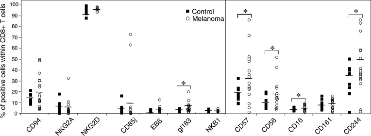
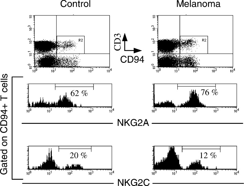
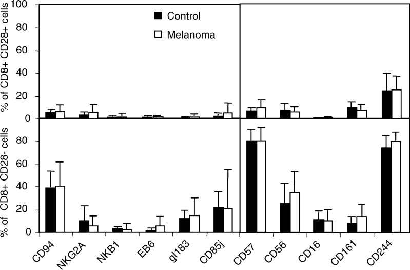
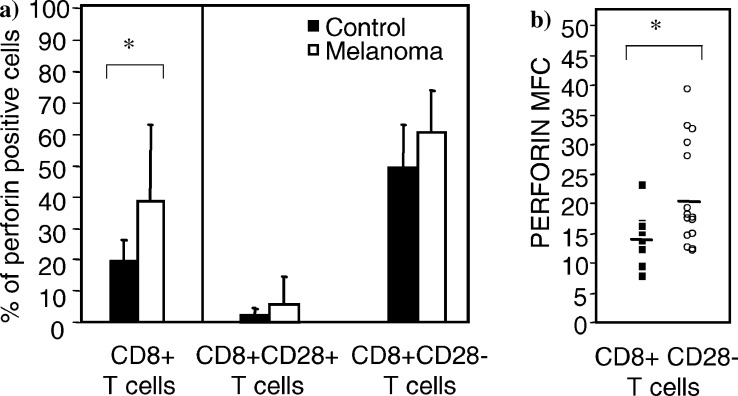
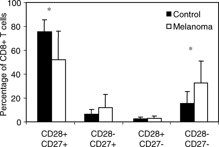
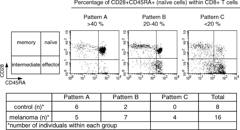
CD8+ T cells can express NK-associated receptors (NKRs) that may regulate their cytolytic function. We have characterized the expression of several NKRs on peripheral blood CD8+ T cells from melanoma patients and compared them to age-matched healthy donors. The analysis performed includes HLA class I specific receptors (KIRs, LILRB1 and CD94/NKG2) and other NK receptors like CD57, CD56 and CD16. Melanoma patients showed a higher variability in the expression of NKRs on circulating CD8+ T cells than age-matched healthy donors. NKR expression on CD8+ T cells from melanoma patients showed a significant increase of KIR2DL2/L3/S2 (mAb gl183), CD244, CD57, CD56 and CD16. We have also found an increase of CD8+ CD28- CD27- T cells in melanoma patients. This subset represents terminally differentiated effector cells expressing CD244 and high levels of perforin. The expression of NKRs was also mainly restricted to this T cell subset. Altogether, circulating CD8+ T cells from melanoma patients display a distinct phenotype characterized by downregulation of costimulatory molecules and higher expression of NKRs. We suggest that the increased expression of NKRs on T cells may contribute to the final outcome of the immune response against melanoma both stimulating or inhibiting activation and differentiation to effector cells. Blocking inhibitory receptor function and enhancing activating receptors may represent new strategies with therapeutic potential against melanoma.
Figures
Similar articles
-
Lymphocyte activation in response to melanoma: interaction of NK-associated receptors and their ligands.Cancer Immunol Immunother. 2007 Jan;56(1):101-9. doi: 10.1007/s00262-006-0141-y. Epub 2006 Feb 17. Cancer Immunol Immunother. 2007. PMID: 16485126 Free PMC article. Review.
-
HLA class I-specific inhibitory receptors in human T lymphocytes: interleukin 15-induced expression of CD94/NKG2A in superantigen- or alloantigen-activated CD8+ T cells.Proc Natl Acad Sci U S A. 1998 Feb 3;95(3):1172-7. doi: 10.1073/pnas.95.3.1172. Proc Natl Acad Sci U S A. 1998. PMID: 9448304 Free PMC article.
-
Increased expression of the natural killer cell inhibitory receptor CD94/NKG2A and CD158b on circulating and lesional T cells in patients with chronic plaque psoriasis.Br J Dermatol. 2006 Aug;155(2):318-24. doi: 10.1111/j.1365-2133.2006.07301.x. Br J Dermatol. 2006. PMID: 16882169
-
CD28-negative cytolytic effector T cells frequently express NK receptors and are present at variable proportions in circulating lymphocytes from healthy donors and melanoma patients.Eur J Immunol. 1999 Jun;29(6):1990-9. doi: 10.1002/(SICI)1521-4141(199906)29:06<1990::AID-IMMU1990>3.0.CO;2-9. Eur J Immunol. 1999. PMID: 10382762
-
Expression of NK-associated receptors on cytotoxic T cells from melanoma patients: a two-edged sword?Cancer Immunol Immunother. 2004 Oct;53(10):911-24. doi: 10.1007/s00262-004-0507-y. Epub 2004 May 4. Cancer Immunol Immunother. 2004. PMID: 15127235 Free PMC article. Review.
Cited by
-
Expression profiling of TCR-engineered T cells demonstrates overexpression of multiple inhibitory receptors in persisting lymphocytes.Blood. 2013 Aug 22;122(8):1399-410. doi: 10.1182/blood-2013-04-495531. Epub 2013 Jul 16. Blood. 2013. PMID: 23861247 Free PMC article.
-
Restored CD8+PD-1+ T Cells Facilitate the Response to Anti-PD-1 for Patients With Pancreatic Ductal Adenocarcinoma.Front Oncol. 2022 Apr 11;12:837560. doi: 10.3389/fonc.2022.837560. eCollection 2022. Front Oncol. 2022. PMID: 35480107 Free PMC article.
-
CD28 negative T cells: is their loss our gain?Am J Transplant. 2014 Nov;14(11):2460-6. doi: 10.1111/ajt.12937. Epub 2014 Oct 16. Am J Transplant. 2014. PMID: 25323029 Free PMC article. Review.
-
CD8+ CD28- and CD8+ CD57+ T cells and their role in health and disease.Immunology. 2011 Sep;134(1):17-32. doi: 10.1111/j.1365-2567.2011.03470.x. Epub 2011 Jun 29. Immunology. 2011. PMID: 21711350 Free PMC article. Review.
-
Analysis of Vδ1 T cells in clinical grade melanoma-infiltrating lymphocytes.Oncoimmunology. 2012 Nov 1;1(8):1297-1304. doi: 10.4161/onci.21659. Oncoimmunology. 2012. PMID: 23243593 Free PMC article.
References
-
- Algarra I, Garcia-Lora A, Cabrera T, Ruiz-Cabello F, Garrido F. The selection of tumor variants with altered expression of classical and nonclassical MHC class I molecules: implications for tumor immune escape. Cancer Immunol Immunother. 2004;53:904. doi: 10.1007/s00262-004-0517-9. - DOI - PMC - PubMed
-
- Andre P, Brunet C, Guia S, Gallais H, Sampol J, Vivier E, Dignat-George F. Differential regulation of killer cell Ig-like receptors and CD94 lectin-like dimers on NK and T lymphocytes from HIV-1-infected individuals. Eur J Immunol. 1999;29:1076. doi: 10.1002/(SICI)1521-4141(199904)29:04<1076::AID-IMMU1076>3.3.CO;2-Q. - DOI - PubMed
-
- Appay V, Dunbar PR, Callan M, Klenerman P, Gillespie GM, Papagno L, Ogg GS, King A, Lechner F, Spina CA, Little S, Havlir DV, Richman DD, Gruener N, Pape G, Waters A, Easterbrook P, Salio M, Cerundolo V, McMichael AJ, Rowland-Jones SL. Memory CD8+ T cells vary in differentiation phenotype in different persistent virus infections. Nat Med. 2002;8:379. doi: 10.1038/nm0402-379. - DOI - PubMed
-
- Bakker AB, Phillips JH, Figdor CG, Lanier LL. Killer cell inhibitory receptors for MHC class I molecules regulate lysis of melanoma cells mediated by NK cells, gamma delta T cells, and antigen-specific CTL. J Immunol. 1998;160:5239. - PubMed
-
- Becker JC, Vetter CS, Schrama D, Brocker EB, thor Straten P. Differential expression of CD28 and CD94/NKG2 on T cells with identical TCR beta variable regions in primary melanoma and sentinel lymph node. Eur J Immunol. 2000;30:3699. doi: 10.1002/1521-4141(200012)30:12<3699::AID-IMMU3699>3.0.CO;2-2. - DOI - PubMed
Publication types
MeSH terms
Substances
LinkOut - more resources
Full Text Sources
Other Literature Sources
Medical
Research Materials
