. 2005 Sep 1;106(5):1544-51.
doi: 10.1182/blood-2004-11-4365. Epub 2005 May 12.

Chimeric NK-receptor-bearing T cells mediate antitumor immunotherapy

Affiliations

Chimeric NK-receptor-bearing T cells mediate antitumor immunotherapy

Tong Zhang et al. Blood. .

Abstract

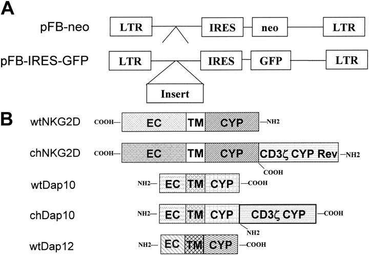
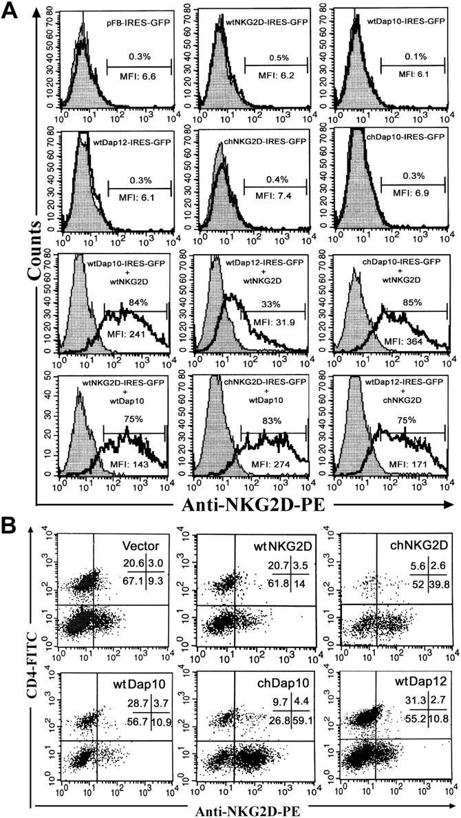
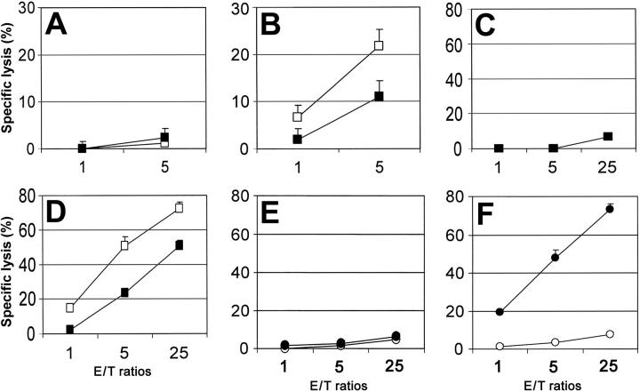
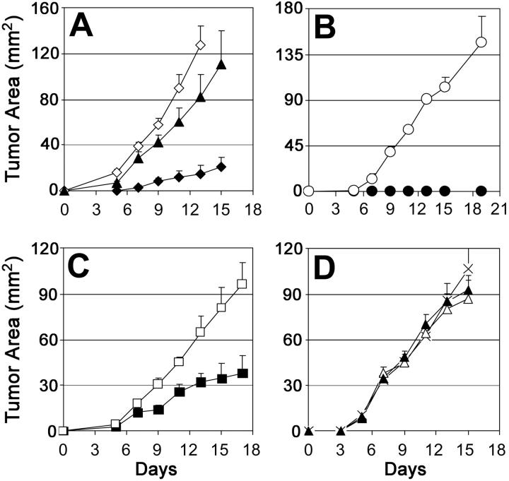
NKG2D is an activating cell-surface receptor expressed on natural killer (NK) cells and some T-cell subsets. Its ligands are primarily expressed on tumor cells. The aim of this study was to determine whether chimeric NK-receptor-bearing T cells would directly kill tumor cells and lead to induction of host immunity against tumors. Chimeric NK receptors were produced by linking NKG2D or DNAX activating protein of 10 kDa (Dap10) to the cytoplasmic portion of the CD3zeta chain. Our results showed that chimeric (ch) NKG2D-bearing T cells responded to NKG2D-ligand-bearing tumor cells (RMA/Rae-1beta, EG7) but not to wild-type tumor cells (RMA). This response was dependent upon ligand expression on the target cells but not on expression of major histocompatibility complex (MHC) molecules, and the response could be blocked by anti-NKG2D antibodies. These T cells produced large amounts of T-helper 1 (Th1) cytokines and proinflammatory chemokines and killed ligand-expressing tumor cells. Adoptive transfer of chNKG2D-bearing T cells inhibited RMA/Rae-1beta tumor growth in vivo. Moreover, mice that had remained tumor-free were resistant to subsequent challenge with the wild-type RMA tumor cells, suggesting the generation of immunity against other tumor antigens. Taken together, our findings indicate that modification of T cells with chimeric NKG2D receptors represents a promising approach for immunotherapy against cancer.


Figures

Figure 1.
Figure 1.
Structure of retroviral constructs. (A) The empty vectors pFB-neo and pFB-IRES-GFP were used in experiments as controls for retrovirus infection. The gene inserts were placed behind the 5′ long terminal repeat (LTR) and upstream of the internal ribosomal entry site (IRES), whereas the marker gene (either neo or GFP) was controlled by the IRES. Schematic diagram of wild-type (wt) and chimeric (ch) proteins are shown in panel B. The extracellular (EC), transmembrane (TM), and cytoplasmic regions (CYP) are indicated. In the chNKG2D, the CD3ζ chain was fused to the N-terminus of the NKG2D molecule in a reverse (Rev) orientation (COOH-terminus > NH2-terminus). In chDap10, the CD3ζ chain was placed downstream of the COOH-terminus of the Dap10 molecule in a normal orientation.
Figure 2.
Figure 2.
chNKG2D and chDap10 molecules express as efficiently as their wild-type counterparts on cell surface. (A) NKG2D expression on Bosc23 cells was evaluated 24 hours after transfection by combination of plasmids containing NKG2D and adaptor genes. Surface expression of NKG2D was determined on the gated GFP+ population using the PE-conjugated anti-NKG2D Ab (unshaded region). Isotype controls are shown in shaded region. The percentage of NKG2D+ cells is indicated. (B) NKG2D expression on B6 T cells 7 days after transduction. NKG2D expression was measured using anti-NKG2D mAbs in combination with anti-CD4 and anti-CD3 mAbs. More than 97% cells are CD3+ T cells (data not shown). CD4 T cells are CD8+ T cells. The dot plots shown were all gated on CD3+ cells. The percentage of cells in each quadrant is indicated. The data are a representative of 6 similar experiments.
Figure 3.
Figure 3.
chNKG2D- or chDap10-modified T cells produce large amounts of Th1 cytokines and proinflammatory chemokines after coincubation with NKG2D ligand–expressing tumor cells. The NKG2D ligand expression on various target cells is shown in panel A. Cells were stained with mouse NKG2D Ig (open curve) or control Ig (shaded curve). Bar labeled M1 designates positive events. For detection of IFN-γ (B), T cells (105) were cocultured with 105 target tumor cells RMA (formula image
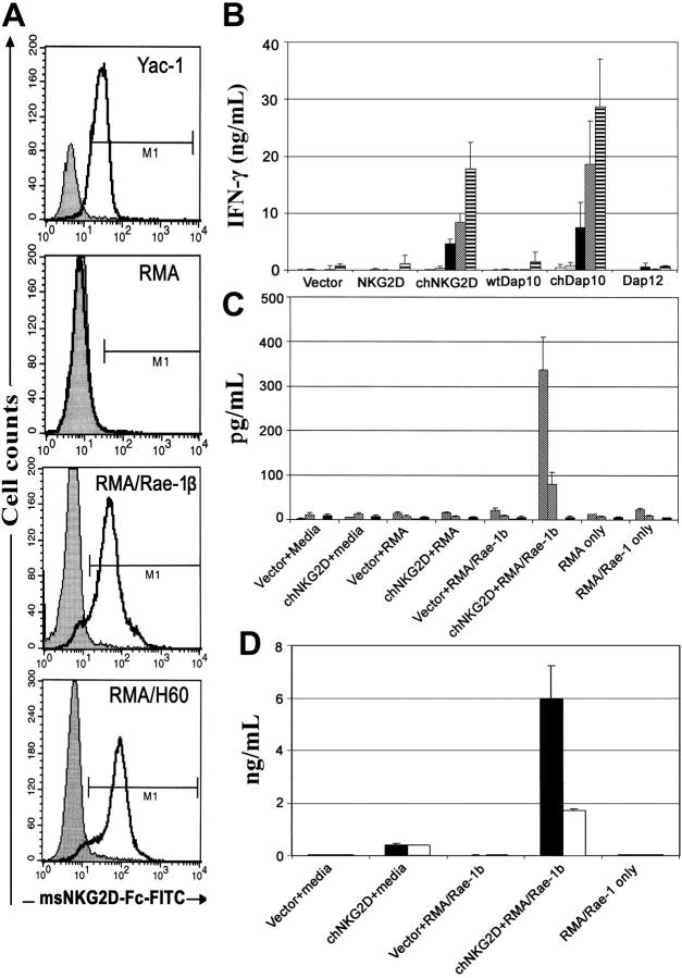
), RMA/Rae-1β (▪), RMA/H60 (formula image), and YAC-1 (formula image) or media alone (□) for 24 hours. Concentrations of IFN-γ in supernatants were determined by ELISA. For detection of other cytokines (C) and chemokines (D), irradiated (100 Gy) tumor cells instead were mixed with T cells for 3 days. Bio-plex assays were performed to measure the levels of GM-CSF (formula image), IL-3 (formula image), IL-5 (formula image), IL-10 (▪) (shown in C), CCL3 (▪), and CCL5 (□) (D).
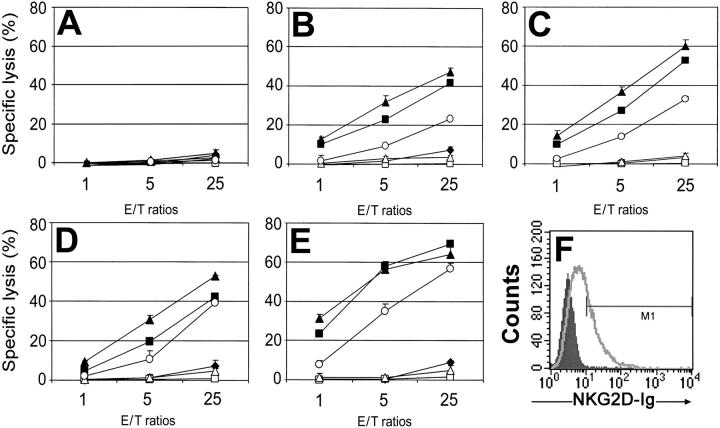
Figure 4.
Figure 4.
Specific lysis of target cells by gene-modified primary T cells. Effector T cells modified with vector only (♦), wtNKG2D (□), chNKG2D (▪), wtDAP10 (▵), chDap10 (▴), or wtDap12 (○) were cocultured with target cells RMA (A), RMA/Rae-1β (B), RMA/H60 (C), YAC-1 (D), or EG7 (E) cells, respectively, at ratios from 1:1 to 25:1 in 4-hour 51Cr release assays. The data are presented as means ± SD and are representative of 3 to 5 independent experiments. (F) The expression of NKG2D ligands on EG7 cells is shown. Cells were stained with mouse NKG2D Ig (open curve) or control Ig (shaded curve).
Figure 5.
Figure 5.
Lytic activity of chNKG2D-bearing T cells is dependent upon NKG2D recognition but not target cell MHC expression. Effector T cells modified with vector only (A,C) or chNKG2D (B,D) were cocultured with target cells RMA/Rae-1β (A-B) or EG7 (C-D) in the presence of anti-NKG2D antibodies (▪) or control antibodies (□), and percentage of specific lysis determined after a 4-hour 51Cr release assay. Effector T cells modified with vector only (○) or chNKG2D (•) were cocultured with RMA-S (E), or RMA-S/Rae-1β (F) cells, respectively, in 4-hour 51Cr release assays. The data are presented as means ± SD and are representative of 2 or 3 independent experiments.
Figure 6.
Figure 6.
Effects of coadministration of chNKG2D-modified T cells with RMA/Rae-1β tumor cells on in vivo tumor growth and generation of host antitumor immunity. (A) chNKG2D (♦)– or vector-only (⋄)–transduced T cells were mixed with RMA/Rae-1β tumor cells at a ratio of 10:1 and injected subcutaneously into the right flank of recipient mice. RMA/Rae-1β cells alone (▴) were also injected as control. The error bars represent SEM. chNKG2D-bearing T cells significantly (P < .05 at days 5-15) suppressed the RMA/Rae-1β tumor growth compared with vector-transduced T cells or tumor alone. (B) Tumor-free mice (•) in the chNKG2D-treated group (A) and age-matched naive mice (○) were challenged with wild-type RMA cells (104) injected subcutaneously into the left flank. A summary of 3 independent experiments is shown. (C) The day before RMA/Rae-1β tumor implantation (day-1), 107 chNKG2D (▪)– or vector-only (□)–transduced T cells were adoptively transferred to mice intravenously. At day 0, tumor cells (105) were implanted subcutaneously at the right flank. The tumor areas are represented as means ± SEM. P < .05 at days 9 to 17. (D) chNKG2D (▴)– or vector-only(▵)–transduced T cells were mixed with wild-type RMA tumor cells at a ratio of 10:1 and injected subcutaneously into B6 mice. RMA cells alone (×) were also injected as control. The error bars represent SEM. There was no significant suppression of RMA tumor growth (P > .05) by chNKG2D-transduced T cells compared with vector-transduced T cells or tumor alone.

References

    1. Rossig C, Brenner MK. Genetic modification of T lymphocytes for adoptive immunotherapy. Mol Ther. 2004;10: 5-18. - PubMed
    1. Sadelain M, Riviere I, Brentjens R. Targeting tumours with genetically enhanced T lymphocytes. Nat Rev Cancer. 2003;3: 35-45. - PubMed
    1. Rosenberg SA. Progress in human tumour immunology and immunotherapy. Nature. 2001;411: 380-384. - PubMed
    1. Ho WY, Blattman JN, Dossett ML, Yee C, Greenberg PD. Adoptive immunotherapy: engineering T cell responses as biologic weapons for tumor mass destruction. Cancer Cell. 2003;3: 431-437. - PubMed
    1. Crowley NJ, Slingluff CL Jr, Darrow TL, Seigler HF. Generation of human autologous melanoma-specific cytotoxic T-cells using HLA-A2-matched allogeneic melanomas. Cancer Res. 1990;50: 492-498. - PubMed

Publication types