. 2005 Jun;79(11):6588-97.
doi: 10.1128/JVI.79.11.6588-6597.2005.

Deletion of M2 gene open reading frames 1 and 2 of human metapneumovirus: effects on RNA synthesis, attenuation, and immunogenicity

Affiliations

Deletion of M2 gene open reading frames 1 and 2 of human metapneumovirus: effects on RNA synthesis, attenuation, and immunogenicity

Ursula J Buchholz et al. J Virol. 2005 Jun.

Abstract

The M2 gene of human metapneumovirus (HMPV) contains two overlapping open reading frames (ORFs), M2-1 and M2-2. The expression of separate M2-1 and M2-2 proteins from these ORFs was confirmed, and recombinant HMPVs were recovered in which expression of M2-1 and M2-2 was ablated individually or together [rdeltaM2-1, rdeltaM2-2, and rdeltaM2(1+2)]. Each M2 mutant virus directed efficient multicycle growth in Vero cells. The ability to recover HMPV lacking M2-1 contrasts with human respiratory syncytial virus, for which M2-1 is an essential transcription factor. Expression of the downstream HMPV M2-2 ORF was not reduced when translation of the upstream M2-1 ORF was silenced, indicating that it is initiated separately. The rdeltaM2-2 mutants exhibited a two- to fivefold increase in the accumulation of mRNA, normalized to the genome template, suggesting that M2-2 has a role in regulating RNA synthesis. Replication and immunogenicity were tested in hamsters. Animals infected intranasally with rdeltaM2-1 or rdeltaM2(1+2) did not have recoverable virus in the lungs or nasal turbinates on days 3 or 5 postinfection and did not develop HMPV-neutralizing serum antibodies or resistance to HMPV challenge. Thus, M2-1 appears to be essential for significant virus replication in vivo. In animals infected with rdeltaM2-2, virus was recovered from only 1 of 12 animals and only in the nasal turbinates on a single day. However, all of the animals developed a high titer of HMPV-neutralizing serum antibodies and were highly protected against challenge with wild-type HMPV. The HMPV rdeltaM2-2 virus is a promising and highly attenuated HMPV vaccine candidate.


Figures

FIG. 1.
FIG. 1.
Overview of the HMPV M2 gene deletion mutants. (A) Map of the HMPV genome (top; scale approximate) and introduced mutations. ORFs are shown as open rectangles with amino acid lengths given below. The locations of restriction sites used for cloning are shown above; the PacI and KpnI sites are naturally occurring, and the NheI and BsiWI sites (boxed) were generated by site-directed mutagenesis (reference and the present study). Also shown is the insertion of an additional gene encoding the enhanced GFP gene constructed as described previously (5): for each mutant, a version was made with and without this added GFP gene (e.g., rgHMPV and rHMPV, respectively). The enlargement shows the PacI-BsiWI fragment that contains the M2 gene, with the ORFs shown as open rectangles and amino acid lengths in parentheses; the gene start and gene end signals are shown as black triangles and rectangles, respectively. Mutations involved in silencing the ORFs include introduced stop codons (stars), deletion of part of the M2-2 ORF or [in the case of ΔM2(1+2)] deletion of the M2 gene in its entirety together with 13 nt of the intergenic (ig) region. The resulting nucleotide lengths of the modified M2 genes are listed in the box. The recombinant viruses were analyzed by RT-PCR. Total cellular RNA was isolated from Vero cells infected with the recombinants. First-strand synthesis was done with a first-strand primer spanning HMPV nt 4656 to 4676. PCR was performed using the same primer and a reverse primer spanning HMPV nt 5529 to 5508, and the products were separated on a 2% agarose gel (right side of panel A) to confirm the presence of the deletions. (B) Details of mutations introduced to silence the M2-1 and M2-2 ORFs. The upstream end of the M2-1 ORF of HMPV (HMPV nt 4724 to 4768) is shown with the mutations involved in ablating expression of M2-1 (top box). The upstream end of the M2-2 ORF overlapping the downstream end of the M2-1 ORF (HMPV nt 5234 to 5288) is shown with mutations involved in ablating expression of M2-2 (bottom box). The wild-type sequence is at the top of each box, and introduced changes are shown below. Translation start and stop codons are in boldface. At the amino acid level, stop codons are shown as stars. The upstream end of the deletion involved in the ΔM2-2 mutant is indicated (Δ). The identities of the encoded proteins are indicated on the right.
FIG. 2.
FIG. 2.
Western blot analysis of HMPV proteins expressed in cells transfected with expression plasmids or infected with wild-type or mutant HMPV. (A) BHK-21 cells that stably express T7 RNA polymerase were transfected with plasmids pT7-N, pT7-P, or pT7-M2-1, which carry the indicated HMPV ORF under control of the T7 promoter (5) (lanes 1 to 3, respectively). Alternatively, Vero cells were infected with rHMPV, rΔM2-1, rΔM2-2, rΔM2(1+2), or HMPV CAN97-83 at an input MOI of 1 PFU/cell or were mock-infected (lanes 4 to 9, respectively). The cells were harvested 72 h after transfection or infection, and lysates were prepared and subjected to sodium dodecyl sulfate-polyacrylamide gel electrophoresis under reducing and denaturing conditions using 4 to 12% gradient gels. After Western blot transfer to nitrocellulose, the membrane was incubated with a rabbit hyperimmune serum against purified HMPV (5, 6) followed by goat anti-rabbit antibodies conjugated with horseradish peroxidase, and the bound antibodies were visualized by chemiluminescence. The N, P, and M2-1 proteins are labeled as well as a species that probably corresponds to M protein. (B) Western blots of cell lysates of Vero cells harvested 72 h after infection with the indicated GFP-expressing recombinants with or without an HA epitope tag fused to the M2-2 protein. The membranes were incubated with hyperimmune serum to HMPV (upper panel) or a mouse monoclonal primary antibody specific to the HA epitope (HA-7; Sigma) (lower panel).
FIG. 3.
FIG. 3.
Northern blot analysis of intracellular HMPV M2 RNA. An aliquot of the Vero cells that had been infected with the panel of wild-type and M2 mutant viruses and harvested for the experiment shown in Fig. 2 was used to isolate total intracellular RNA. Cells were infected with the rHMPV, rΔM2-1, rΔM2-2, rΔM2(1+2), or HMPV CAN97-83 viruses or were mock infected (lanes 1 to 6, respectively). The RNA was electrophoresed on a 1% agarose gel in the presence of 0.44 M formaldehyde, transferred to nylon membrane, and hybridized with [α-32P]-labeled double-stranded DNA probes specific for HMPV M2. Radioactivity was detected with a phosphorimager. Monocistronic M2 mRNA and the M2-SH and M2-SH-G readthrough mRNAs (5, 6) are identified on the right.
FIG. 4.
FIG. 4.
Northern blot time course analysis of the accumulation of intracellular HMPV RNA. Replicate cultures of Vero cells were infected with rgHMPV or the indicated M2 mutants and harvested 12, 24, 36, 48, and 72 h later. Total intracellular RNA was isolated, separated as described in the legend to Fig. 3, and analyzed by Northern blot hybridization using single-stranded negative-sense (upper panel) and positive-sense (lower panel) RNA probes synthesized in vitro from F cDNA using SP6 and T7 RNA polymerase, respectively.
FIG. 5.
FIG. 5.
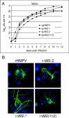
(A) Comparison of the multistep growth kinetics of rgHMPV and the M2 mutants. Vero cells were infected at an MOI of 0.01 PFU/cell with rgHMPV, rgΔM2-1, rgΔM2-2, or rgΔM2(1+2). At 24-h intervals, medium samples (0.5 ml of the 2-ml overlay) were taken and replaced by an equivalent volume of fresh medium containing 5 μg/ml of trypsin. The samples were analyzed by plaque assay and immunostaining with anti-HMPV antiserum. Each time point was represented by two wells, and each virus titration was done in duplicate. Means are shown. (B) Indirect immunofluorescence assay of Vero cells 4 days following infection at an MOI of 0.03 with the indicated recombinants. Cells were fixed with 4% buffered paraformaldehyde and permeabilized with 1% Triton X-100. HMPV antigens were visualized with a rabbit hyperimmune serum to HMPV, followed by an Alexa488-conjugated goat anti-rabbit antibody (Molecular Probes). Nuclear chromatin staining (blue) was performed with 4′,6-diamidino-2-phenylindole (DAPI; Sigma).

References

    1. Ahmadian, G., P. Chambers, and A. J. Easton. 1999. Detection and characterization of proteins encoded by the second ORF of the M2 gene of pneumoviruses. J. Gen. Virol. 80:2011-2016. - PubMed
    1. Ahmadian, G., J. S. Randhawa, and A. J. Easton. 2000. Expression of the ORF-2 protein of the human respiratory syncytial virus M2 gene is initiated by a ribosomal termination-dependent reinitiation mechanism. EMBO J. 19:2681-2689. - PMC - PubMed
    1. Bermingham, A., and P. L. Collins. 1999. The M2-2 protein of human respiratory syncytial virus is a regulatory factor involved in the balance between RNA replication and transcription. Proc. Natl. Acad. Sci. USA 96:11259-11264. - PMC - PubMed
    1. Biacchesi, S., M. H. Skiadopoulos, G. Boivin, C. T. Hanson, B. R. Murphy, P. L. Collins, and U. J. Buchholz. 2003. Genetic diversity between human metapneumovirus subgroups. Virology 315:1-9. - PubMed
    1. Biacchesi, S., M. H. Skiadopoulos, K. C. Tran, B. R. Murphy, P. L. Collins, and U. J. Buchholz. 2004. Recovery of human metapneumovirus from cDNA: optimization of growth in vitro and expression of additional genes. Virology 321:247-259. - PubMed

MeSH terms

LinkOut - more resources