. 2005 Jun;79(11):6620-30.
doi: 10.1128/JVI.79.11.6620-6630.2005.

Selective replication of coronavirus genomes that express nucleocapsid protein

Affiliations

Selective replication of coronavirus genomes that express nucleocapsid protein

Barbara Schelle et al. J Virol. 2005 Jun.

Abstract

The coronavirus nucleocapsid (N) protein is a structural protein that forms a ribonucleoprotein complex with genomic RNA. In addition to its structural role, it has been described as an RNA-binding protein that might be involved in coronavirus RNA synthesis. Here, we report a reverse genetic approach to elucidate the role of N in coronavirus replication and transcription. We found that human coronavirus 229E (HCoV-229E) vector RNAs that lack the N gene were greatly impaired in their ability to replicate, whereas the transcription of subgenomic mRNA from these vectors was easily detectable. In contrast, vector RNAs encoding a functional N protein were able to carry out both replication and transcription. Furthermore, modification of the transcription signal required for the synthesis of N protein mRNAs in the HCoV-229E genome resulted in the selective replication of genomes that are able to express the N protein. This genetic evidence leads us to conclude that at least one coronavirus structural protein, the N protein, is involved in coronavirus replication.


Figures
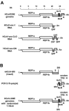
FIG. 1.
FIG. 1.
Structure of recombinant HCoV-229E and vector RNAs. (A) The structural relationship and sizes of HCoV-229E and vector RNAs used in this study are shown. Open reading frames are indicated as boxes designated by encoded gene products. L, leader RNA sequence; An, poly(A) sequence. (B) The structure of recombinant HCoV-229E genomes with a randomized N gene TRS is shown (bottom). Also shown are the cDNAs that have been ligated in vitro (at the SfiI site) to produce a full-length HCoV-229E cDNA template for in vitro transcription.
FIG. 2.
FIG. 2.
Transfection of vector RNAs. Vector RNA HCoV-vec-GN or HCoV-vec-1 (each, 15 μg) was transfected into BHK-21 cells with or without synthetic mRNAs (5 μg) encoding the HCoV-229E, PEDV, or MHV N proteins or a truncated β-galactosidase protein as indicated. Three days posttransfection, the percentage of green fluorescent cells was analyzed by flow cytometry. Each column represents the mean value of three independent experiments.
FIG. 3.
FIG. 3.
Northern blot analysis. Poly(A)-containing RNA was isolated from HCoV-229E-infected MRC-5 cells (lane 1), BHK-21 cells (lane 6), BHK-21 cells that have been transfected with HCoV-229E N protein mRNA and HCoV-vec-1 RNA (lanes 7 and 10), BHK-21 cells that have been transfected with HCoV-229E N protein mRNA and HCoV-vec-GN RNA (lane 11), or BHK-21 cells that have been transfected with HCoV-229E N protein mRNA, HCoV-vec-CLG RNA, and HCoV-229E genomic RNA (lane 12). The RNA was analyzed by Northern blotting as described in Materials and Methods. Lane 2, no RNA; lanes 3 and 8, 1 ng of in vitro-transcribed HCoV-vec-1 RNA; lanes 4 and 9, 1 ng of in vitro-transcribed HCoV-vec-CLG RNA; lane 5, 1 ng of in vitro-transcribed HCoV-229E N protein mRNA. Lanes 8, 9, and 10 represent lanes 3, 4, and 7, respectively, after prolonged autoradiography. Genomic and subgenomic RNAs of HCoV-229E (RNA1 to -7) and HCoV-229E vector RNAs (arrows) are indicated.
FIG. 4.
FIG. 4.
Firefly luciferase reporter protein analysis in BHK-HCoV-N cells. (A) Western blot analysis of BHK-HCoV-N cells. Each lane corresponds to a cytoplasmic lysate derived from 2 × 10⁵ BHK-HCoV-N cells that were cultivated for 2 days in medium without or with 0.01, 0.1, or 1 μg/ml doxycyclin (Dox), as indicated. For comparison, a Western blot analysis of cytoplamic lysate derived from 2 × 10⁵ HCoV-229E-infected MRC-5 cells is shown. (B)A total of 15 μg of HCoV-vec-CLG vector RNA was transfected into 107 BHK-HCoV-N cells. After transfection, the cells were split into four wells containing medium without or with 0.01, 0.1, or 1 μg/ml Dox. Luciferase activity was analyzed 3 days posttransfection. Results are shown for three independent experiments. In each experiment, luciferase activity of transfected BHK-HCoV-N cells that were cultivated without Dox were set as 1 relative unit. Bars indicate 95% confidence intervals.
FIG. 5.
FIG. 5.
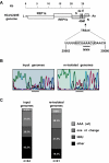
Sequencing analysis of recombinant HCoV-229E genomes with a randomized N gene TRS. (A) HCoV-229E nt 25663 to 25680 containing the core sequence of the N gene TRS (boxed) are shown together with the structure and size of the HCoV-229E genome. The HCoV-229E nt 25671AAA25673 within the TRS-N core sequence (underlined) was changed to a random sequence indicated as NNN. (B) An RT-PCR sequencing analysis of in vitro-transcribed HCoV-229E-based genomes with randomized HCoV-229E nt 25671 to 25673 is shown (input genomes) (left). These genomes were used for transfection of BHK-21 cells. Three days later, the poly(A)-containing RNA was isolated and analyzed by RT-PCR sequencing (reisolated genome) (right). Sequencing results are shown for HCoV-229E nt 25663 to 25680, and the region corresponding to the randomized sequence is underlined. (C) RT-PCR products obtained from input genomes or reisolated genomes were cloned and sequenced. The analysis comprised 44 individual plasmid clones corresponding to input genomes and 41 individual plasmid clones corresponding to reisolated genomes. On the basis of the sequence determined at randomized nt 25671 to 25673, the recombinant genomes were placed in one of four groups: group 1, recombinant genomes with the HCoV-229E wild-type sequence (AAA); group 2, recombinant genomes with a 1-nt change compared to the TRS-N or leader-TRS sequence; group 3, recombinant genomes containing a U nucleotide at HCoV-229E position 25673 (NNU); group 4, sequences that do not match to groups 1 to 3. The percentages of each group in the population of input and reisolated genomes are indicated.
FIG. 6.
FIG. 6.
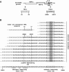
Sequence analysis of subgenomic N protein mRNAs. (A) The structure and size of the recombinant HCoV-229E genome with randomized nt 25671 to 25673 are shown together with the core sequences of the leader TRS and TRS-N. Numbers indicate nucleotide positions within the HCoV-229E genome. (B) The leader-body junction sequences of subgenomic N protein mRNAs are shown, determined after leader-body-specific RT-PCR sequencing analysis. Leader and body-derived nucleotides are shown in gray and black letters, respectively. For each subgenomic N protein mRNA, the region of the TRS-N core sequence (box) and the sequence corresponding to the randomized nt 25671 to 25673 (shaded in gray) are indicated. For comparison, HCoV-229E nt 55 to 78 encoding part of the leader RNA and downstream sequences (top left) and the HCoV-229E nt 25623 to 25684 containing the TRS-N and flanking regions are shown (bottom). Also highlighted are two stretches of 8 nt encoded downstream of the leader TRS (nt 70 to 77) and upstream of the TRS-N (nt 25668 to 25674) that might be involved in base pairing during discontinuous extension (see Discussion). Alternative leader-body fusion sites are underlined.

References

    1. Almazan, F., C. Galan, and L. Enjuanes. 2004. The nucleoprotein is required for efficient coronavirus genome replication. J. Virol. 78:12683-12688. - PMC - PubMed
    1. Baric, R. S., G. W. Nelson, J. O. Fleming, R. J. Deans, J. G. Keck, N. Casteel, and S. A. Stohlman. 1988. Interactions between coronavirus nucleocapsid protein and viral RNAs: implications for viral transcription. J. Virol. 62:4280-4287. - PMC - PubMed
    1. Barr, J. N., S. P. Whelan, and G. W. Wertz. 2002. Transcriptional control of the RNA-dependent RNA polymerase of vesicular stomatitis virus. Biochim. Biophys. Acta 1577:337-353. - PubMed
    1. Blumberg, B. M., M. Leppert, and D. Kolakofsky. 1981. Interaction of VSV leader RNA and nucleocapsid protein may control VSV genome replication. Cell 23:837-845. - PubMed
    1. Bol, J. F. 1999. Alfalfa mosaic virus and ilarviruses: involvement of coat protein in multiple steps of the replication cycle. J. Gen. Virol. 80:1089-1102. - PubMed

Publication types

MeSH terms

LinkOut - more resources