. 2005 Jun;79(11):6723-31.
doi: 10.1128/JVI.79.11.6723-6731.2005.

Interaction of tSNARE syntaxin 18 with the papillomavirus minor capsid protein mediates infection

Affiliations

Interaction of tSNARE syntaxin 18 with the papillomavirus minor capsid protein mediates infection

Ioannis Bossis et al. J Virol. 2005 Jun.

Abstract

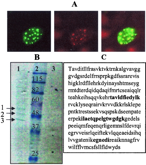
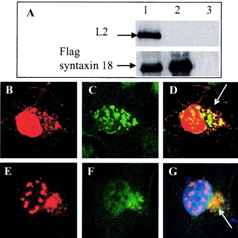
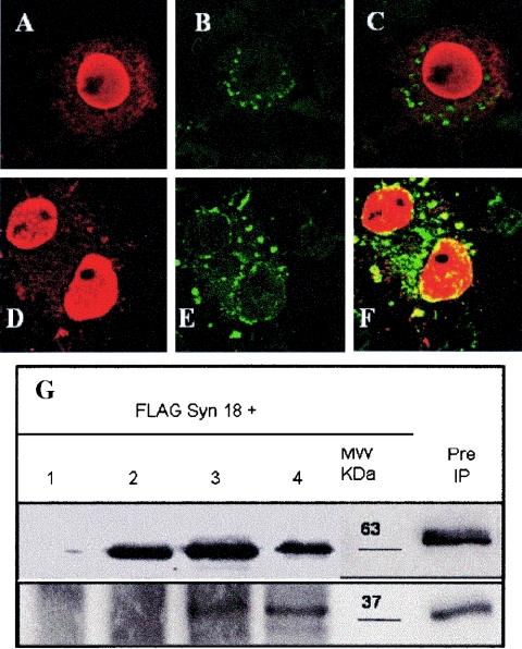
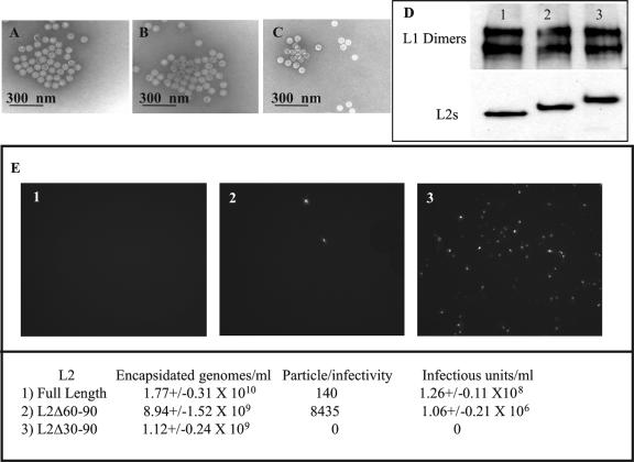
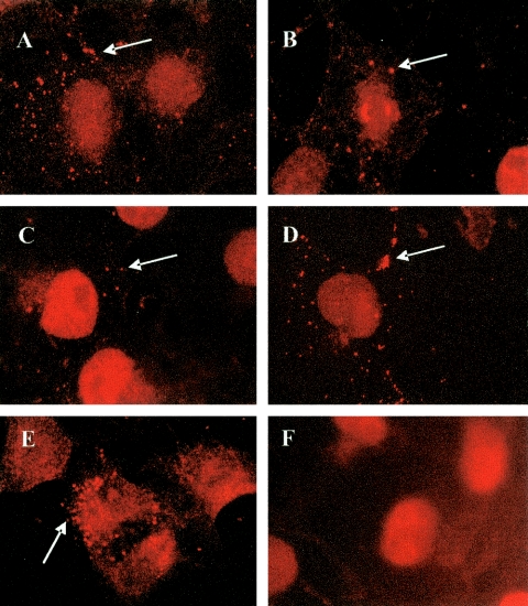
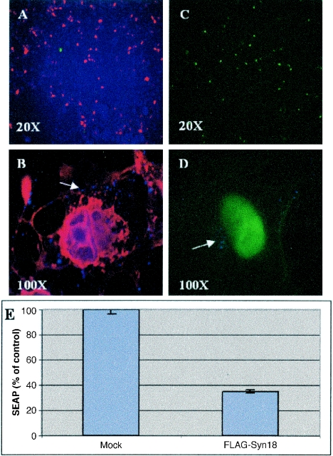
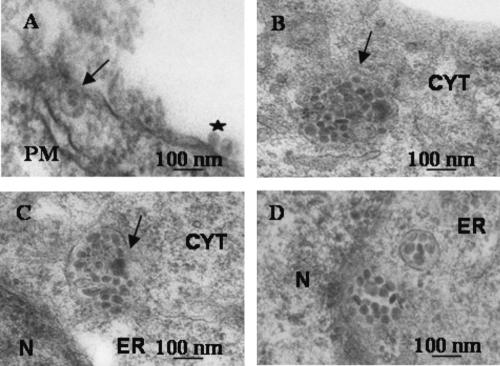
The papillomavirus capsid mediates binding to the cell surface and passage of the virion to the perinuclear region during infection. To better understand how the virus traffics across the cell, we sought to identify cellular proteins that bind to the minor capsid protein L2. We have identified syntaxin 18 as a protein that interacts with bovine papillomavirus type 1 (BPV1) L2. Syntaxin 18 is a target membrane-associated soluble N-ethylmaleimide-sensitive factor-attachment protein receptor (tSNARE) that resides in the endoplasmic reticulum (ER). The ectopic expression of FLAG-tagged syntaxin 18, which disrupts ER trafficking, blocked BPV1 pseudovirion infection. Furthermore, the expression of FLAG-syntaxin 18 prevented the passage of BPV1 pseudovirions to the perinuclear region that is consistent with the ER. Genetic studies identified a highly conserved L2 domain, DKILK, comprising residues 40 to 44 that mediated BPV1 trafficking through the ER during infection via an interaction with the tSNARE syntaxin 18. Mutations within the DKILK motif of L2 that did not significantly impact virion morphogenesis or binding at the cell surface prevented the L2 interaction with syntaxin 18 and disrupted BPV1 infection.


Figures

FIG. 1.
FIG. 1.
Identifying the interaction of BPV1 L2 with syntaxin 18. (A) ECTAP BPV1L2 is properly distributed within cells. COS-7 cells were transfected with ECTAP BPV1L2 for 24 h. The left panel shows staining for BPV1 L2, the middle panel shows staining for PML, and the right panel shows a merged image. (B) Coomassie blue-stained gel of TAP extracts from ECTAP BPV1L2-transfected COS-7 cells (lane 1) and ECTAP-transfected control cells (lane 3). Molecular weight markers are shown in lane 2. The proteins labeled 1, 2, and 3 in lane 1 were identified as L2, actin, and syntaxin 18, respectively. No proteins were seen in lane 3. (C) Peptide matches to the syntaxin 18 sequence are shown in bold.
FIG. 2.
FIG. 2.
Syntaxin 18 and BPV1 L2 coimmunoprecipitate and colocalize. (A) COS-7 cells were cotransfected with FLAG-syntaxin 18 and hBPV L2 (lane 1), transfected with FLAG-syntaxin 18 (lane 2), or transfected with hBPV1 L2 (lane 3). Cells were harvested after 24 h in RIPA buffer, and samples were immunoprecipitated with a FLAG antibody. The top panel was blotted with mouse monoclonal anti-L2 (clone C6), and the bottom panel was blotted with a rabbit polyclonal anti-syntaxin 18 antibody. L2 coimmunoprecipitated with FLAG-syntaxin 18 (lane 1) and did not immunoprecipitate with the FLAG antibody (lane 3). (B to D) COS-7 cells cotransfected with FLAG-syntaxin 18 and hBPV1 L2 were fixed and stained after 24 h for BPV1 L2 (B) and for FLAG (C). Overlapping fluorescence is indicated by an arrow (D). (E to G) COS-7 cells transfected with hBPV1 L2 were fixed and stained after 24 h for BPV1 L2 (E) and for endogenous syntaxin 18 (F). Overlapping fluorescence is indicated by an arrow (G).
FIG. 3.
FIG. 3.
BPV1 L2 residues 30 to 60 mediate the interaction with syntaxin 18. COS-7 cells were transfected with FLAG-syntaxin 18 and either L2 Δ30-90 (A to C) or L2 Δ60-90 (D to F). The cells were fixed and stained after 24 h with rabbit anti-BPV1 L2 (A and D) or a mouse anti-FLAG M2 antibody (B and E). The merged images show no overlapping of syntaxin 18 with L2 Δ30-90 (C) and do show overlapping with L2 Δ60-90 (F). (G) FLAG-syntaxin 18 was cotransfected with the pcDNA3 vector (lane 1), L2 Δ30-90 (lane 2), L2 Δ60-90 (lane 3), or hBPV1 L2 (lane 4). The molecular size marker shows the protein sizes in kilodaltons (kDa), and preimmunoprecipitated full-length L2 and FLAG-syntaxin 18 are also shown. Immunoprecipitation was performed with the anti-L2 monoclonal C6 antibody and Western blotting was performed with a rabbit anti-BPV1 L2 antibody (top), with a mouse anti-FLAG antibody (bottom) used on the same blot. The results showed that L2 Δ60-90 and full-length BPV1 L2 coimmunoprecipitated with FLAG-syntaxin 18, whereas L2 Δ30-90 did not. The L2 C6 antibody did not immunoprecipitate FLAG-syntaxin 18 (lane 1, bottom panel).
FIG. 4.
FIG. 4.
Role of hBPV1 L2 residues 30 to 90 in viral production and infection. The electron microscopic images of virions generated with L2 Δ30-90 (A), L2 Δ60-90 (B), and hBPV1 L2 (C) seem indistinguishable. (D) Denatured Nycodenz-purified virions (lane 1, Δ30-90; lane 2, Δ60-90; lane 3, BPV1 L2) run in a 10% SDS-PAGE gel were blotted for L1 (upper panel) and L2 (lower panel). (E) COS-7 cells (3 × 10⁶) were incubated with pseudovirions generated with L2 Δ30-90 (panel 1), L2 Δ60-90 (panel 2), and BPV1 L2 (panel 3). Infected cells fluoresced green due to GFP transduction. The levels of genome encapsidation, particle-to-infectivity ratios, and infectious units per ml were determined and are summarized in the bottom panel.
FIG. 5.
FIG. 5.
BPV1 pseudovirion infection. COS-7 cells were infected with BPV pseudovirions generated with the following L2s: Δ30-90 (A), Δ31-44 (B), Δ41-54 (C), D40A (D), and ANS (E). Virions are shown in orange after staining with the 5B6 antibody (arrows). (F) Control, uninfected COS-7 cells stained with 5B6 show no punctate fluorescence indicative of pseudovirions.
FIG. 6.
FIG. 6.
FLAG-syntaxin 18 prevents BPV1 pseudovirion infection. (A and B) COS-7 cells transfected with FLAG-syntaxin 18 12 h before being infected with particles containing wild-type L2. Cells were immunostained for FLAG-syntaxin 18 (red) and GFP was visualized (green). Over 40% of cells were transfected with syntaxin 18, and few cells were infected. (C and D) Vector-transfected COS-7 cells infected with BPV1 L2 pseudovirions had 50% GFP transduction at 24 h. Panels B and D show merged images of GFP fluorescence, pseudovirion staining with the 5B6 antibody (blue dots; arrows), and FLAG-syntaxin 18 staining with the M2 antibody (red). (E) Summary of three experiments showing the level of SEAP expression on cells infected with SEAP-containing wild-type L2 pseudoviruses. FLAG-syntaxin 18-transfected cells had 30% ± 3.4% expression compared to mock-transfected cells after infection.
FIG. 7.
FIG. 7.
Ultrastructural analysis of BPV1 pseudovirion infection. Cells were preincubated at 4°C for 2 h with virions before being warmed to 37°C. Samples were obtained 5 min (A), 2 h (B), and 7 h (C and D) after the warming step. (A) A virion is bound at the plasma membrane (PM; star), and internalization into a vesicle is occurring (arrow). (B) A virion-filled vesicle has pinched off from the plasma membrane into the cytoplasm (arrow). (C) The ER and a virus-filled vesicle membrane are fusing (arrow). (D) Viruses are found free floating in the ER or perinuclear (N, nucleus) region and in a formed vesicle.

References

    1. Becker, K. A., L. Florin, C. Sapp, G. G. Maul, and M. Sapp. 2004. Nuclear localization but not PML protein is required for incorporation of the papillomavirus minor capsid protein L2 into virus-like particles. J. Virol. 78:1121-1128. - PMC - PubMed
    1. Becker, K. A., L. Florin, C. Sapp, and M. Sapp. 2003. Dissection of human papillomavirus type 33 L2 domains involved in nuclear domains (ND) 10 homing and reorganization. Virology 314:161-167. - PubMed
    1. Booy, F. P., R. B. Roden, H. L. Greenstone, J. T. Schiller, and B. L. Trus. 1998. Two antibodies that neutralize papillomavirus by different mechanisms show distinct binding patterns at 13 A resolution. J. Mol. Biol. 281:95-106. - PubMed
    1. Bosch, F. X., A. Lorincz, N. Munoz, C. J. Meijer, and K. V. Shah. 2002. The causal relation between human papillomavirus and cervical cancer. J. Clin. Pathol. 55:244-265. - PMC - PubMed
    1. Brobst, D. F., and G. C. Dulac. 1969. Meningeal tumors induced in calves with the bovine cutaneous papilloma virus. Pathol. Vet. 6:135-145. - PubMed

Publication types

MeSH terms

LinkOut - more resources