. 2005 Jun;79(11):7207-16.
doi: 10.1128/JVI.79.11.7207-7216.2005.

Poliovirus proteins induce membrane association of GTPase ADP-ribosylation factor

Affiliations

Poliovirus proteins induce membrane association of GTPase ADP-ribosylation factor

George A Belov et al. J Virol. 2005 Jun.

Abstract

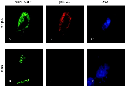
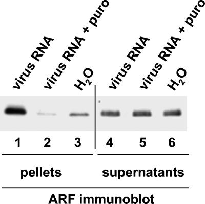
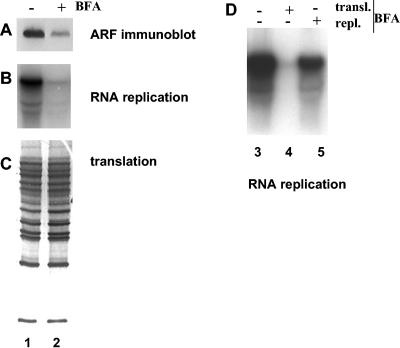
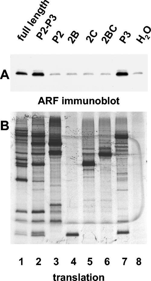
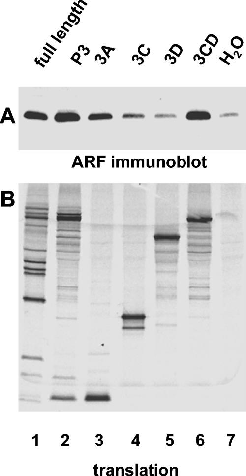
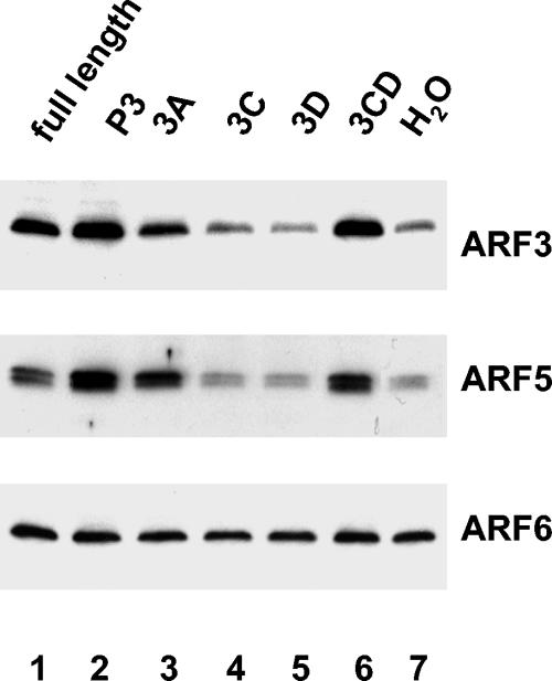
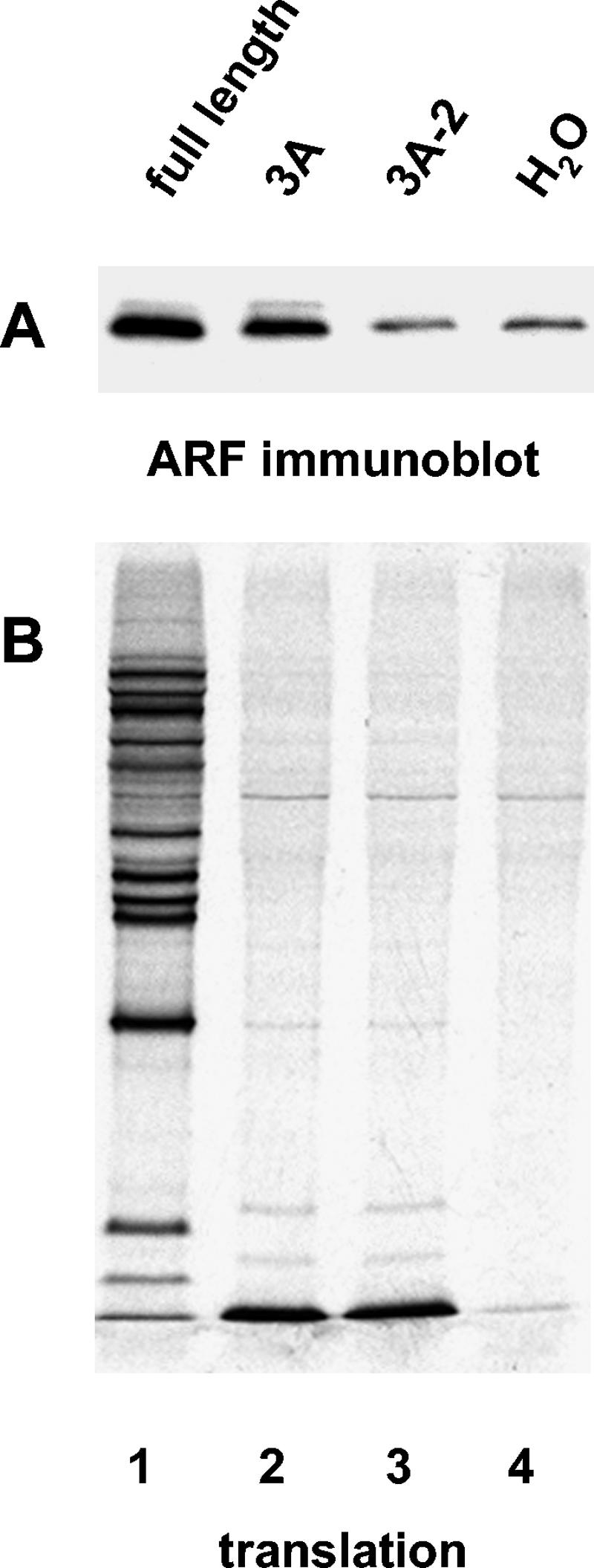
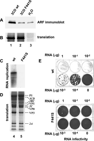
Poliovirus infection results in the disintegration of intracellular membrane structures and formation of specific vesicles that serve as sites for replication of viral RNA. The mechanism of membrane rearrangement has not been clearly defined. Replication of poliovirus is sensitive to brefeldin A (BFA), a fungal metabolite known to prevent normal function of the ADP-ribosylation factor (ARF) family of small GTPases. During normal membrane trafficking in uninfected cells, ARFs are involved in vesicle formation from different intracellular sites through interaction with numerous regulatory and coat proteins as well as in regulation of phospholipase D activity and cytoskeleton modifications. We demonstrate here that ARFs 3 and 5, but not ARF6, are translocated to membranes in HeLa cell extracts that are engaged in translation of poliovirus RNA. The accumulation of ARFs on membranes correlates with active replication of poliovirus RNA in vitro, whereas ARF translocation to membranes does not occur in the presence of BFA. ARF translocation can be induced independently by synthesis of poliovirus 3A or 3CD proteins, and we describe mutations that abolished this activity. In infected HeLa cells, an ARF1-enhanced green fluorescent protein fusion redistributes from Golgi stacks to the perinuclear region, where poliovirus RNA replication occurs. Taken together, the data suggest an involvement of ARF in poliovirus RNA replication.


Figures

FIG. 1.
FIG. 1.
Intracellular relocation of ARF-1-EGFP protein following infection of HeLa cells with poliovirus. Cells were transfected with pARF1-EGFP 24 h prior to infection with poliovirus (A, B, and C) or mock infection (D, E, and F). Six hours postinfection, cells were examined for EGFP fluorescence (A and D) or stained with anti-polio 2C antibodies (B and E) and Hoechst 33342 for nuclear DNA (C and F).
FIG. 2.
FIG. 2.
Translocation of ARF to membranes induced by translation of poliovirus proteins in vitro. Poliovirus RNA was translated in HeLa S10 extracts for 3.5 h. Reactions were fractionated by centrifugation, and total pellets (lanes 1 to 3) and one-tenth of the total volume of supernatants (lanes 4 to 6) were analyzed by immunoblotting with anti-ARF antibody. Lanes: 1 and 4, fractions from reactions translating viral RNA; 2 and 5, fractions from reactions in which viral RNA translation was inhibited by 1 mg/ml of puromycin; 3 and 6, fractions from reactions incubated with no RNA.
FIG. 3.
FIG. 3.
Inhibition of ARF translocation and viral RNA replication by BFA. Viral RNA was translated in HeLa S10 extracts for 3.5 h. (A to C) Duplicate reactions were fractionated by centrifugation, and the pellets were analyzed by immunoblotting with anti-ARF antibody (A) and assayed for viral RNA replication (B) as described in Materials and Methods. Translation was monitored by SDS-PAGE of [35S]methionine-labeled proteins synthesized during the translation step (C). Lane 1, no BFA was present during either translation or replication steps; lane 2, BFA (80 μg/ml) was present only during the translation step. (D) RNA replication after BFA addition during translation or replication steps. Lane 3, no BFA was present during either translation or replication steps; lane 4, BFA (80 μg/ml) was present only during the translation step; lane 5, BFA (80 μg/ml) was present only during the replication step.
FIG. 4.
FIG. 4.
Determinants specifying ARF translocation map to the P3 region of the poliovirus genome. RNA transcripts were translated in HeLa S10 extracts for 3.5 h. (A) Reactions were fractionated by centrifugation, and the pellets were analyzed by immunoblotting with anti-ARF antibody. (B) Translation of each transcript was monitored by SDS-PAGE of [35S]methionine-labeled proteins. The coding region of the RNA transcripts used to program the translation reaction is indicated above each lane.
FIG. 5.
FIG. 5.
Poliovirus proteins 3A and 3CD can induce ARF translocation independently. RNA transcripts were translated in HeLa S10 extracts for 3.5 h. (A) Reactions were fractionated by centrifugation, and the pellets were analyzed by immunoblotting with anti-ARF antibody. (B) Translation was monitored by SDS-PAGE of [35S]methionine-labeled proteins. The coding region of each RNA transcript used to program the translation reaction is indicated above each lane.
FIG. 6.
FIG. 6.
Activation of individual ARFs induced by translation of poliovirus-specific transcripts. RNA transcripts were translated in HeLa S10 extracts for 3.5 h. Reactions were fractionated by centrifugation, and the pellets were analyzed by immunoblotting. The membrane shown in Fig. 5 was stripped and probed sequentially with specific antibodies against ARF3, ARF5, or ARF6.
FIG. 7.
FIG. 7.
Mutant 3A-2 does not activate ARF. RNA transcripts were translated in HeLa S10 extracts for 3.5 h. (A) Reactions were fractionated by centrifugation, and the pellets were analyzed by immunoblotting with anti-ARF antibodies. (B) Translation was monitored by SDS-PAGE of [35S]methionine-labeled proteins. The coding region of each RNA transcript used to program the translation reaction is indicated above each lane.
FIG. 8.
FIG. 8.
Mutation 3CD F441S prevents ARF translocation and inhibits viral RNA replication in vitro and virus growth in vivo. Wild-type (lane 1), mutant (lane 2), or no (lane 3) 3CD RNA transcripts were translated in HeLa S10 extracts for 3.5 h. (A) Reactions were fractionated by centrifugation, and the pellets were analyzed by immunoblotting with anti-ARF antibody. (B) [35S]methionine-labeled translation products. (C) Wild-type (lane 4) or mutant (lane 5) full-length genomic transcripts were translated in HeLa S10 extracts for 3.5 h. Reactions were fractionated by centrifugation, and the pellets were assayed for viral RNA replication. (D) [35S]methionine-labeled translation products. (E) HeLa cell monolayers were transfected with serial dilutions of wild-type or mutant full-length genomic transcripts. Cells were covered after transfection with agarose-solidified medium and stained 48 h later with crystal violet.

References

    1. Aldabe, R., and L. Carrasco. 1995. Induction of membrane proliferation by poliovirus proteins 2C and 2BC. Biochem. Biophys. Res. Commun. 206:64-76. - PubMed
    1. Altan-Bonnet, N., R. D. Phair, R. S. Polishchuk, R. Weigert, and J. Lippincott-Schwartz. 2003. A role for Arf1 in mitotic Golgi disassembly, chromosome segregation, and cytokinesis. Proc. Natl. Acad. Sci. USA 100:13314-13319. - PMC - PubMed
    1. Balch, W. E., R. A. Kahn, and R. Schwaninger. 1992. ADP-ribosylation factor is required for vesicular trafficking between the endoplasmic reticulum and the cis-Golgi compartment. J. Biol. Chem. 267:13053-13061. - PubMed
    1. Barco, A., and L. Carrasco. 1995. A human virus protein, poliovirus protein 2BC, induces membrane proliferation and blocks the exocytic pathway in the yeast Saccharomyces cerevisiae. EMBO J. 14:3349-3364. - PMC - PubMed
    1. Barco, A., and L. Carrasco. 1998. Identification of regions of poliovirus 2BC protein that are involved in cytotoxicity. J. Virol. 72:3560-3570. - PMC - PubMed

MeSH terms

LinkOut - more resources