

Rapid and fully automated visualization of subdural electrodes in the presurgical evaluation of epilepsy patients
Affiliations
- PMID: 15891163
- PMCID: PMC8158601
Item in Clipboard
Rapid and fully automated visualization of subdural electrodes in the presurgical evaluation of epilepsy patients
AJNR Am J Neuroradiol.
2005 May.
Abstract
For rapid visualization of subdural electrodes with respect to cortical and subcortical structures, we describe a novel and fully automated method based on coregistration, normalization, optional cerebellum masking, and volume rendering of 3D MR imaging data taken before and after implantation. The key step employs the skull-stripped preimplantation image as a mask to also remove the skull in the postimplantation image. The extracted brain is presented in 3D with the electrodes directly visible by their susceptibility artifacts. Compared with alternative methods, ours is based on freely available software and does not require manual intervention.
Figures
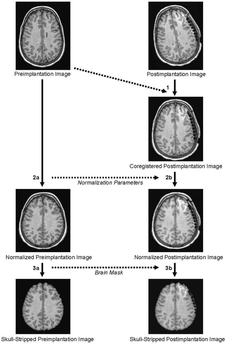
The image data processing is fully automated and consists of the following steps: 1) coregistration of the postimplantation image to the preimplantation image; 2A) normalization of the preimplantation image; 2B) normalization of the postimplantation image based on the transformation parameters derived from the normalization of the preimplantation image; 3A) brain extraction in the preimplantation image; and 3B) brain extraction in the coregistered postimplantation image by using the skull-stripped preimplantation image as a mask.
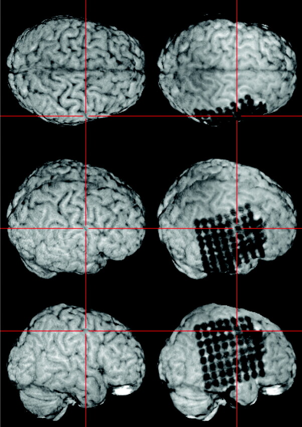
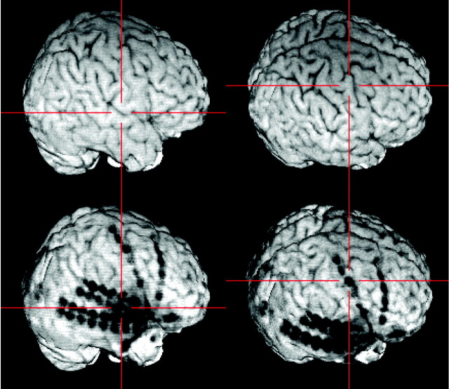
The resulting skull-stripped postimplantation image is visualized within MRIcro. A simultaneous examination of the skull-stripped preimplantation and postimplantation 3D MR images in two yoked MRIcro application windows gives an excellent overview of the exact location of each electrode contact. The viewer is able to grasp the electrodes’ spatial relation to the sulcal pattern of the brain and to anatomic landmarks (e.g., the central sulcus as pointed out here by the crosshairs).
Subdural strip and double strip electrodes and their relationship to the Sylvian fissure (left) and to the precentral gyrus (right).
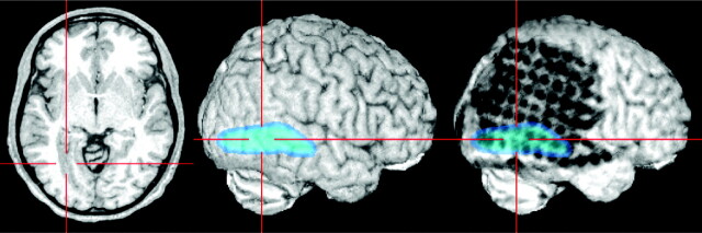
Lesions beneath the grid or the brain surface (here polymicrogyria) can be highlighted by loading as color-coded overlays and subsequent semitransparent volume rendering. The individual position of each electrode contact with respect to the presumed epileptogenic lesion can be exactly depicted by using yoked coregistered planar MR images alongside the 3D MR imaging data.
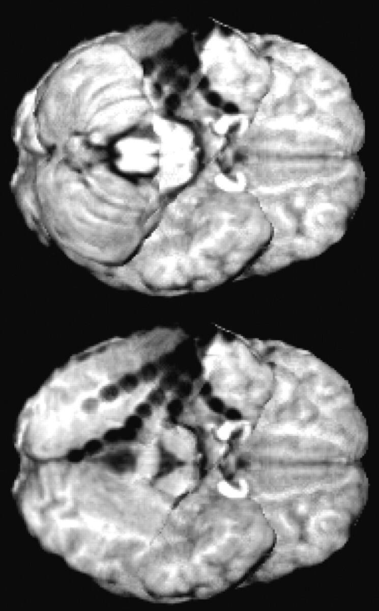
To allow for optimal visualization of electrodes on the posterior basal surface of the brain, the brain extraction (see Fig 1, step 3B) is optionally combined with masking the cerebellum in the skull-stripped postimplantation image.
References
-
- Behrens E, Zentner J, van Roost D, et al. Subdural and depth electrodes in the presurgical evaluation of epilepsy. Acta Neurochir (Wien) 1994;128:84–87 - PubMed
-
- Winkler PA, Vollmar C, Krishnan KG, et al. Usefulness of 3-D reconstructed images of the human cerebral cortex for localization of subdural electrodes in epilepsy surgery. Epilepsy Res 2000;41:169–178 - PubMed
-
- Noordmans HJ, van Rijen PC, van Veelen CW, et al. Localization of implanted EEG electrodes in a virtual-reality environment. Comput Aided Surg 2001;6:241–258 - PubMed
-
- Wellmer J, von Oertzen J, Schaller C, et al. Digital photography and 3D MRI-based multimodal imaging for individualized planning of resective neocortical epilepsy surgery. Epilepsia 2002;43:1543–1550 - PubMed
MeSH terms
LinkOut - more resources
Full Text Sources
Other Literature Sources
Medical