

Relationship between brain tissue oxygen tension and CT perfusion: feasibility and initial results
- PMID: 15891166
- PMCID: PMC8158594
Relationship between brain tissue oxygen tension and CT perfusion: feasibility and initial results
Abstract
Background and purpose: Monitoring of intraparenchymal brain tissue oxygen tension (P(br)O(2)) is an emerging tool in neurocritical care. The purpose of this study was to determine if there is a relationship between CT perfusion (CTP) imaging parameters and P(br)O(2).
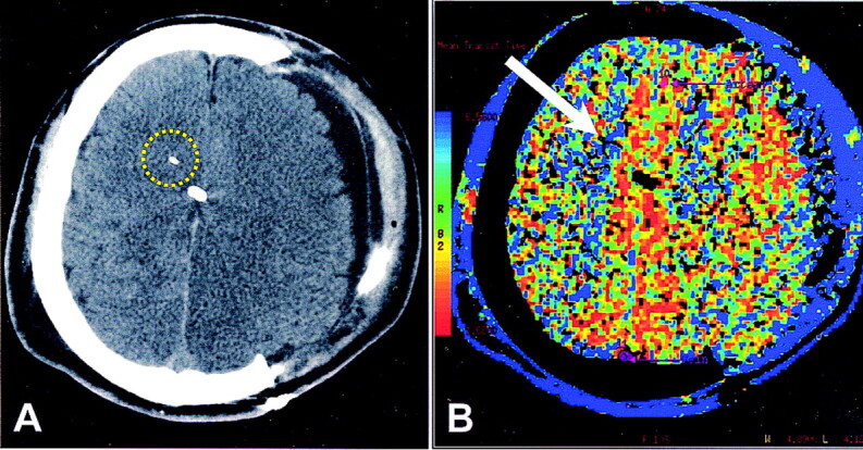
Methods: Nineteen patients underwent continuous P(br)O(2) monitoring with probes placed to target white matter in the cerebral hemisphere. Twenty-two CTP studies were performed at the level of the oxygen electrode, as identified on concurrent nonenhanced CT. CTP analysis software was used to measure mean transit time (MTT) and cerebral blood volume (CBV) and to derive cerebral blood flow (CBF) for a region of interest (ROI) surrounding the oxygen probe. For correlation, P(br)O(2) levels and other physiologic parameters were recorded at the time of CTP.
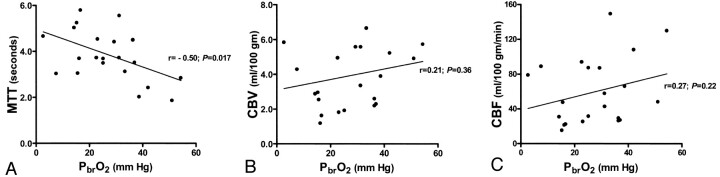
Results: P(br)O(2) values at the time of CTP were 2.7-54.4 mm Hg, MTT was 1.86-5.79 seconds, CBV was 1.18-8.76 mL/100 g, and CBF was 15.2-149.2 mL/100 g/min. MTT but not CBV or CBF was correlated with P(br)O(2) (r = -0.50, P = .017). MTT, CBV, or CBF were not correlated with other physiologic parameters, including mean arterial pressure, cerebral perfusion pressure, intracranial pressure, and fraction of inspired oxygen. On multivariable analysis, only P(br)O(2) was independently associated with MTT.
Conclusion: CTP assessment of ROI surrounding an oxygen probe in the intraparenchymal brain tissue is feasible and showed a significant correlation between P(br)O(2) and MTT. Further studies are warranted to determine the role of CTP in assessing acute brain injury and whether it can be used to prospectively identify brain regions at risk for tissue hypoxia that should be targeted for advanced neuromonitoring.
Figures
References
-
- Goodman JC, Valadka AB, Gopinath SP, Uzura M, Robertson CS. Extracellular lactate and glucose alterations in the brain after head injury measured by microdialysis. Crit Care Med 1999;27:1965–1973 - PubMed
-
- Gopinath SP, Valadka AB, Uzura M, Robertson CS. Comparison of jugular venous oxygen saturation and brain tissue PO2 as monitors of cerebral ischemia after head injury. Crit Care Med 1999;27:2337–2345 - PubMed
-
- Sarrafzadeh AS, Sakowitz OW, Kiening KL, Benndorf G, Lanksch WR, Unterberg AW. Bedside microdialysis: a tool to monitor cerebral metabolism in subarachnoid hemorrhage patients? Crit Care Med 2002;30:1062–1070 - PubMed
-
- Sarrafzadeh AS, Sakowitz OW, Callsen TA, Lanksch WR, Unterberg AW. Detection of secondary insults by brain tissue pO2 and bedside microdialysis in severe head injury. Acta Neurochir Suppl 2002;81:319–321 - PubMed
Publication types
MeSH terms
Substances
Grants and funding
LinkOut - more resources
Full Text Sources
Medical