Comparative Study
. 2005 May;26(5):1128-35.

Trigeminal ganglion and its divisions: detailed anatomic MR imaging with contrast-enhanced 3D constructive interference in the steady state sequences

Affiliations
Comparative Study

Trigeminal ganglion and its divisions: detailed anatomic MR imaging with contrast-enhanced 3D constructive interference in the steady state sequences

Indra Yousry et al. AJNR Am J Neuroradiol. 2005 May.

Abstract

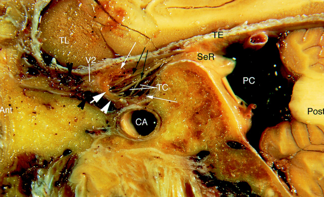
Background and purpose: Visualization of the trigeminal system is important for imaging diagnosis but technically challenging. We assessed how well the trigeminal ganglion, its rootlets, and its branches (V1, V2, and V3) are depicted on three high-resolution pulse sequences.

Methods: Twenty-two patients (44 sides) underwent nonenhanced 3D constructive interference in the steady state (CISS) MR imaging. Two of these patients and another 20 (44 sides) also underwent contrast-enhanced 3D CISS and contrast-enhanced 3D time-of-flight (TOF) MR angiographic (MRA) imaging. Appearances of the ganglion, sinus ganglii, ganglion lip, and sensory and motor rootlets in the Meckel cave were assessed.

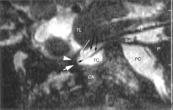
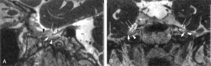
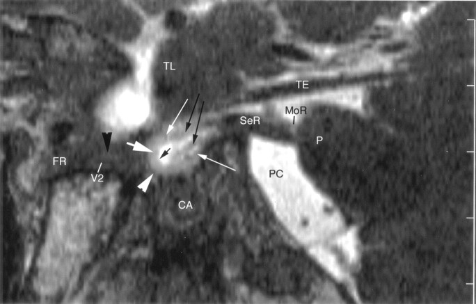
Results: The trigeminal ganglion was shown on enhanced 3D CISS images in all cases, on nonenhanced 3D CISS images in 77.3%, and on enhanced 3D TOF MRA images in 92.9%. Sinus ganglia and lips were demonstrated on 98% of enhanced 3D CISS images. Sensory rootlets were depicted with all 3D CISS sequences but no 3D TOF sequences. V1, V2, and V3 were displayed with all enhanced 3D TOF MRA sequences, 79.5-100% of enhanced 3D CISS sequences, and 0-50% of nonenhanced 3D CISS sequences.

Conclusion: The enhanced 3D CISS sequence was best for displaying the trigeminal ganglion, sinus ganglii, and sinus lips, whereas the enhanced 3D TOF sequence best displayed the emerging V1, V2, and V3 roots. The enhanced 3D CISS sequence was most useful. Complete MR imaging evaluation of the trigeminal ganglion and roots is best performed by using enhanced 3D CISS and enhanced 3D TOF MRA sequences.


Figures

F<sc>ig</sc> 1.
Fig 1.
Sagittal cryomicrotomic section through the Meckel cave in the plane of the trigeminal nerves was obtained from the right medial aspect of a cadaveric specimen from a 74-year-old woman. Small portion of the sensory root (SeR) is seen at its entry into the cave. Trigeminal ganglion (thick white arrow) is at the anterior aspect of the cave, and anterior margin of the ganglion adheres to the dural wall (white arrowhead). Multiple sensory rootlets (long black arrows) arise from the concave medial surface of the ganglion (sinus ganglii) (short black arrow) to course in the trigeminal cistern (TC). Superior and inferior lips of the ganglion (thin white arrows) are particularly well displayed. Portion of the maxillary nerve (V2) shown in this plane is surrounded by venous channels (black arrowheads) along the inferior border of the cavernous sinus. Ant = anterior, CA = carotid artery, PC = prepontine cistern, Post = posterior, TE = cerebellar tentorium, TL = temporal lobe.
F<sc>ig</sc> 2.
Fig 2.
Sagittal nonenhanced 3D CISS image through the right side of the Meckel cave in a 58-year-old woman. Sensory root (SeR) is shown from its apparent origin at the pons (P) to the entrance of the cave. Ganglion (thick white arrow) at the anterior aspect of the cave is difficult to identify, and anterior margin of the trigeminal ganglion cannot be distinguished from the dural wall (arrowhead) of the cave. Only the superior lip of the ganglion (thin white arrow) is well defined. One smaller and one larger sensory rootlet (long black arrows) arise from the sinus ganglii (short black arrow) and course in the trigeminal cistern (TC). Maxillary nerve is not depicted at the inferior border of the cavernous sinus. CA = carotid artery, PC = prepontine cistern, TE = cerebellar tentorium, TL = temporal lobe.
F<sc>ig</sc> 3.
Fig 3.
Enhanced 3D CISS images through the right side of the Meckel cave in a 62-year-old woman. TL = temporal lobe. A, Sagittal image shows a small portion of the sensory root (SeR) at its entry into the cave. Trigeminal ganglion (thick white arrow) and its lips (thin white arrows) are shown at the anterior end of the cave. Anterior margin of the ganglion is clearly differentiated from the dural wall (white arrowhead) of the cave. Two sensory rootlets (long black arrows) emerge from the sinus ganglii (short black arrow) and pass through the trigeminal cistern. Maxillary nerve (V2) courses along the inferior wall of the cavernous sinus, surrounded by enhancing venous channels (black arrowhead). CA = carotid artery, FR = foramen rotundum, PC = prepontine cistern, TE = cerebellar tentorium. B, Coronal image shows the cave bilaterally. Trigeminal ganglion (thick white arrows) lies along the anteroinferior border of the cave. Less-enhancing dura (arrowheads) is clearly differentiated from the ganglion. Multiple, small sensory rootlets (thin white arrows) are seen in the trigeminal cistern (TC). Mandibular nerve (V3) is partly shown on the right side in the foramen ovale (FO).
F<sc>ig</sc> 4.
Fig 4.
Sagittal enhanced 3D CISS image through the right side of the Meckel cave in a 54-year-old man. Sensory root (SeR) is displayed from its apparent origin at the pons (P) to the entrance of the cave. Trigeminal ganglion (thick white arrow) is at the anteroinferior border of the cave. Anterior margin of the enhancing ganglion is clearly distinguished from the less-enhancing dura (white arrowhead) of the cave. Lips of the ganglion (thin white arrows) define the sinus ganglii (short black arrow) from which small sensory rootlets (long black arrows) arise. Maxillary nerve (V2) is depicted along the inferior border of the cavernous sinus, surrounded by venous channels (black arrowhead) en route to the foramen rotundum (FR). CA = carotid artery, MoR = motor root of the trigeminal nerve at its point of exit, PC = prepontine cistern, TE = cerebellar tentorium, TL = temporal lobe.
F<sc>ig</sc> 5.
Fig 5.
Sagittal enhanced 3D TOF image through the right side of the Meckel cave in a 53-year-old-man. Enhancing ganglion (thick white arrow) is shown at the anteroinferior margin of the cave, in continuity with the dural wall (white arrowhead). Superior and inferior lips of the ganglion (thin white arrows) are well depicted. No sensory rootlets are seen in the trigeminal cistern (TC). Ophthalmic nerve (V1) and maxillary nerve (V2) are hypointense, linear structures surrounded by strongly enhancing venous channels (black arrowheads) in the lateral wall and along the inferior border, respectively, of the cavernous sinus. V2 enters the foramen rotundum (FR), while V1 passes to the superior orbital fissure (SOF). CA = carotid artery, PC = prepontine cistern, TE = cerebellar tentorium, TL = temporal lobe.

References

    1. Leblanc A. Encephalo-Peripheral Nervous System. Vascularisation, Anatomy, Imaging. 2nd ed. Berlin: Springer-Verlag;2001. :81–210
    1. Kehrli P, Maillot C, Wolff M-J Anatomy and embryology of the trigeminal nerve and its branches in the parasellar area. Neurol Res 1997;19:57–65 - PubMed
    1. Chui M, Tucker W, Hudson A, Bayer N. High resolution CT of Meckel’s cave. Neuroradiology 1985;27:403–409 - PubMed
    1. Rubinstein D, Stears RLG, Stears JC. Trigeminal nerve and ganglion in the Meckel cave: appearance at the CT and MR imaging. Radiology 1994;193:155–159 - PubMed
    1. Ferner H. On the anatomy of the intracranial segments of the trigeminal nerve [in German]. Z Anat Entwicklungsgesch 1948;114:108–122 - PubMed

Publication types