Comparative Study
. 2005 May;26(5):1178-85.

Perfusion CT for head and neck tumors: pilot study

Affiliations
Comparative Study

Perfusion CT for head and neck tumors: pilot study

Zoran Rumboldt et al. AJNR Am J Neuroradiol. 2005 May.

Abstract

Background and purpose: Differentiation of malignant from benign head and neck lesions is often very difficult on imaging studies, especially in patients with treated cancer. We evaluated the feasibility and reproducibility of perfusion CT (CTP) after enhanced head and neck CT and attempted to differentiate benign from malignant processes.

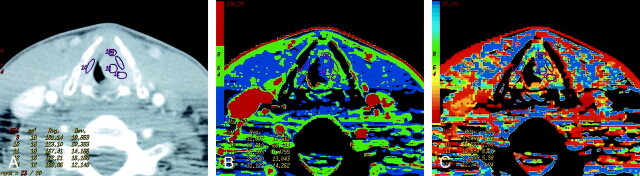
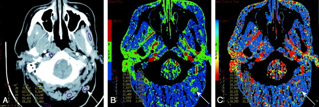
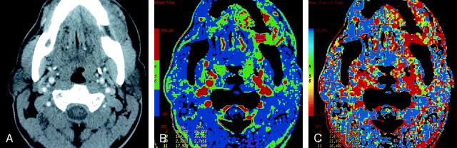
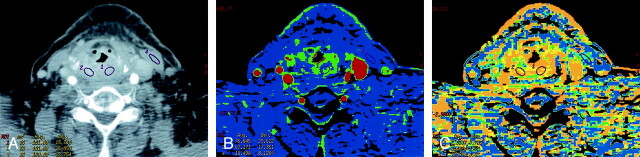
Methods: CTP was attempted in 17 patients after head and neck contrast-enhanced CT. Data were postprocessed by using deconvolution-based perfusion analysis. Ipsilateral and contralateral internal, external, and common carotid arteries were used as arterial input vessels. Postprocessing-generated maps showed mean transit time (MTT), blood volume, blood flow, and capillary permeability surface product. Two readers independently placed regions of interest through the primary site, salivary glands, thyroid gland, paraspinous muscles, muscles of mastication, sternocleidomastoid muscle, base of tongue, and subcutaneous fat. One reader repeated the measurements on separate dates. Data were statistically analyzed, and histologic specimens were obtained.

Results: CTP was not possible in four patients, and one was lost to follow-up. Of the remaining 12, five had cancer, and seven had benign processes. We found no significant interreader or intrareader differences and no significant difference between various input vessels. Differentiation between malignant and nonmalignant lesions was most reliable by using MTTs. Measurements were comparable to those in the literature.

Conclusion: CTP after enhanced head and neck CT is feasible, except perhaps at the laryngeal level. It appears to be reader independent and reproducible regardless of the input vessel. CTP shows promise in distinguishing benign and malignant processes, primarily by means of MTTs.


Figures

F<sc>ig</sc> 1.
Fig 1.
Patient 8 with atypical vascular proliferation of the vocal cord. A, Contrast-enhanced neck CT scan shows a mass in the left true vocal cord. ROIs 9 and 11–13 are in the lesion, and ROI 10 is in the contralateral vocal cord. B, CTP functional map shows a relatively low blood flow of 26–32 mL/100 g/min in the lesion in the left vocal cord. Right vocal cord has blood flow of 26 mL/100 g/min. Scale from red to blue corresponds to decreasing flow. C, CTP functional map shows that the lesion has a relatively long MTT of 6.9–11.6 seconds. Scale from red to blue corresponds to increasing MTT.
F<sc>ig</sc> 2.
Fig 2.
Patient 3 with surgically treated acinic cell cancer of the parotid gland. A, Rater 1. Contrast-enhanced neck CT scan shows a mass in the suboccipital subcutaneous fat (arrow) that was a suspected metastatic lymph node. Histology showed fat necrosis and chronic inflammation. ROIs are placed on the lesion (1) and on the masseter (3) and paraspinal (5) muscles. B, Rater 2. CTP functional map shows relatively low blood flow of 24.9 mL/100 g/min in the lesion (arrow, ROI 1) and shows ROIs in the masseter (4) and paraspinal (8 and 9) muscles. Scale from red to blue corresponds to decreasing flow. C, Rater 2. CTP functional map shows that the lesion (arrow, ROI 1) has a relatively long MTT of 10.5 seconds and shows ROIs in the masseter (4) and paraspinal (8 and 9) muscles. Scale from red to blue corresponds to increasing MTT.
F<sc>ig</sc> 3.
Fig 3.
Patient 12 with primary gingival squamous cell cancer. A, Contrast-enhanced neck CT scan shows a mass on both sides of the left mandibular body (arrows). B, Lesion is more conspicuous on the CTP functional map, which reveals a high blood flow of 96–144 mL/100 g/min. Scale from red to blue corresponds to decreasing flow. C, CTP functional map shows that the lesion has a short MTT of 2.1–5.4 seconds. Scale from red to blue corresponds to increasing MTT. The ROIs were placed within the lesion ( and 9), paraspinal muscles (10), and left parotid gland (11).
F<sc>ig</sc> 4.
Fig 4.
Patient 13 with recurrent laryngeal squamous cell cancer previously treated with radiation. A, Contrast-enhanced neck CT scan shows thickening and diffuse enhancement of supralaryngeal structures. ROIs 1 and 2 are placed bilaterally in the abnormality. Both sampled sites were positive for recurrence on histology. ROI 4 is in the left sternocleidomastoid muscle. B, CTP functional map reveals areas of increased perfusion with blood flows of 67 and 77 mL/100 g/min. Scale from red to blue corresponds to decreasing flow. C, CTP functional map shows short MTTs of 2.5 and 3.4 seconds in the abnormality. Scale from red to blue corresponds to decreasing MTT.

References

    1. Miles KA, Griffiths MR. Perfusion CT: a worthwhile enhancement? Br J Radiol 2003;76:220–231 - PubMed
    1. Cenic A, Nabavi DG, Craen RA, Gelb AW, Lee TY. Dynamic CT measurement of cerebral blood flow: a validation study. AJNR Am J Neuroradiol 1999;20:63–73 - PubMed
    1. Miles KA. Perfusion CT for the assessment of tumour vascularity: which protocol [Suppl]? Br J Radiol 2003;76:S36–S42 - PubMed
    1. Wintermark M, Thiran JP, Maeder P, Schnyder P, Meuli R. Simultaneous measurement of regional cerebral blood flow by perfusion CT and stable Xenon CT: a validation study. AJNR Am J Neuroradiol 2001;22:905–914 - PMC - PubMed
    1. Cenic A, Nabavi DG, Craen RA, Gelb AW, Lee TY. A CT method to measure hemodynamics in brain tumors: validation and application of cerebral blood flow maps. AJNR Am J Neuroradiol 2002;21:462–470 - PMC - PubMed

Publication types