

Case Reports
Cerebral arteriovenous fistulas induced by dural arteriovenous shunts
Affiliations
- PMID: 15891194
- PMCID: PMC8158602
Item in Clipboard
Case Reports
Cerebral arteriovenous fistulas induced by dural arteriovenous shunts
AJNR Am J Neuroradiol.
2005 May.
Abstract
Dural arteriovenous shunts and pial arteriovenous fistulas are uncommonly associated. Their etiology, pathogenesis, and natural history are still unclear and are likely different. We present three cases of high-flow dural arteriovenous shunts associated with pial arteriovenous fistulas and discuss their pathogenesis, anatomic association, and angioarchitecture. We propose that venous steal effect in the dural sinus secondary to the high-flow dural arteriovenous shunt induced the pial arteriovenous fistulas. Treatment of the high-flow dural arteriovenous shunts and the induced pial arteriovenous fistulas are discussed.
Figures
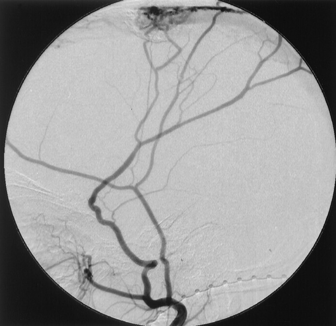
Right external carotid angiogram of case 3 shows the DAVSs at the superior sagittal sinus.
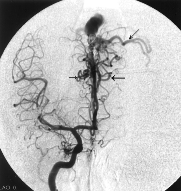
Frontal-view right internal carotid angiogram of case 1 shows pial AVFs (horizontal arrows) and cortical venous reflux (oblique arrow).
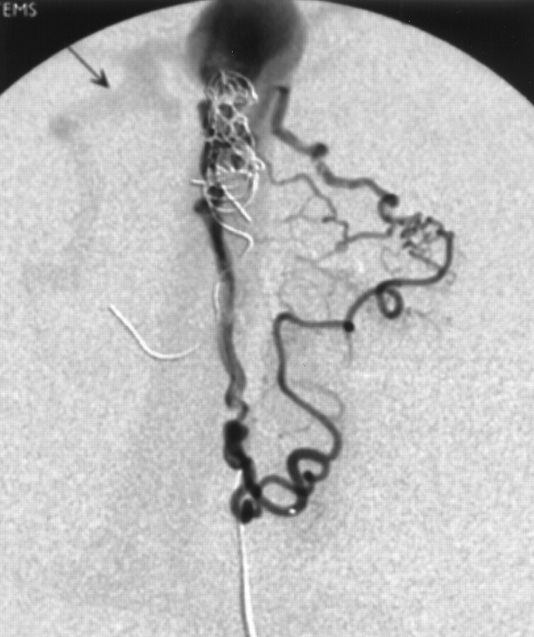
Selective injection of the left anterior cerebral branch of case 1 shows detailed angioarchitecture of the induced pial AVF. Reflux into the cortical vein in the right side is indicated (arrow).
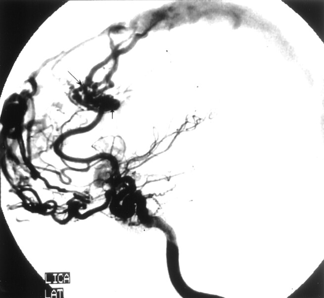
Left internal carotid angiogram of case two shows induced pial AVFs draining to the superior sagittal sinus (large arrow). A flow-related aneurysm has formed (vertical arrow). Reflux into cortical vein is noted (small arrow).
Similar articles
-
Angioarchitecture of arteriovenous fistulas at the craniocervical junction: a multicenter cohort study of 54 patients.J Neurosurg. 2018 Jun;128(6):1839-1849. doi: 10.3171/2017.3.JNS163048. Epub 2017 Sep 1. J Neurosurg. 2018. PMID: 28862546
-
Multiple dural arteriovenous fistulas of the cranium and spine.AJNR Am J Neuroradiol. 1991 May-Jun;12(3):441-5. AJNR Am J Neuroradiol. 1991. PMID: 2058490 Free PMC article.
-
Acquired pial arteriovenous fistula following cerebral vein thrombosis.Stroke. 1999 Nov;30(11):2487-90. doi: 10.1161/01.str.30.11.2487. Stroke. 1999. PMID: 10548689
-
A case of multiple pial arteriovenous fistulas associated with dural arteriovenous fistula.J Neurosurg. 2008 Dec;109(6):1103-7. doi: 10.3171/JNS.2008.109.12.1103. J Neurosurg. 2008. PMID: 19035726 Review.
-
Endovascular treatment of cerebral dural and pial arteriovenous fistulas.Neuroimaging Clin N Am. 2013 Nov;23(4):625-36. doi: 10.1016/j.nic.2013.03.010. Epub 2013 May 16. Neuroimaging Clin N Am. 2013. PMID: 24156854 Review.
Cited by
-
Anterior Cranial Fossa Dural Arteriovenous Fistula with Pial Arterial Supply.Asian J Neurosurg. 2020 Feb 25;15(1):176-179. doi: 10.4103/ajns.AJNS_288_19. eCollection 2020 Jan-Mar. Asian J Neurosurg. 2020. PMID: 32181197 Free PMC article.
-
Variations and management for patients with craniocervical junction arteriovenous fistulas: Comparison of dural, radicular, and epidural arteriovenous fistulas.Surg Neurol Int. 2021 Aug 16;12:411. doi: 10.25259/SNI_557_2021. eCollection 2021. Surg Neurol Int. 2021. PMID: 34513175 Free PMC article.
-
Endovascular Approach to Concurrent Anterior Cranial Fossa Dural AVF and Concurrent Flow-Related Ophthalmic Artery Aneurysm: A Case Study.Asian J Neurosurg. 2024 Nov 25;20(1):183-189. doi: 10.1055/s-0044-1792162. eCollection 2025 Mar. Asian J Neurosurg. 2024. PMID: 40041600 Free PMC article.
-
The role of surgical disconnection for posterior fossa pial arteriovenous fistulas and dural fistulas with pial supply: an illustrative case series.Neurosurg Rev. 2024 Apr 25;47(1):189. doi: 10.1007/s10143-024-02407-y. Neurosurg Rev. 2024. PMID: 38658425
-
Gamma Knife radiosurgery for the treatment of intracranial dural arteriovenous fistulas.Interv Neuroradiol. 2017 Apr;23(2):211-220. doi: 10.1177/1591019916683689. Epub 2016 Dec 16. Interv Neuroradiol. 2017. PMID: 28156167 Free PMC article.
References
-
- SchlachterL, Fleicher A, Faria M, et al. Multifocal intracranial arteriovenous malformations. Neurosurgery 1980;7:440–444 - PubMed
-
- Willinsky RA, Lasjaunias P, Ter Brugge K, Burrows P. Multiple cerebral arteriovenous malformations: review of our experience from 203 patients with cerebral vascular lesions. Neuroradiology 1990;32:207–210 - PubMed
-
- Vilela P, Ter Brugge K, Willinsky R. Association of distinct intracranial pial and dural arteriovenous shunts. Neuroradiology 2001;43:770–777 - PubMed
-
- Taylor CL, Selman WR, Ratcheson RA. Steal affecting the central nervous system. Neurosurg 2002;4:679–688 - PubMed
-
- Garcia-Monaco R, Rodesch G, ter Brugge K, et al. Multifocal dural arteriovenous shunts in children. Childs Nerv Syst 1991;7:425–431 - PubMed
Publication types
MeSH terms
LinkOut - more resources
Full Text Sources