

Proton MR spectroscopic imaging depicts diffuse axonal injury in children with traumatic brain injury
- PMID: 15891197
- PMCID: PMC8158612
Proton MR spectroscopic imaging depicts diffuse axonal injury in children with traumatic brain injury
Abstract
Background and purpose: Diffuse axonal injury (DAI) after traumatic brain injury (TBI) is important in patient assessment and prognosis, yet they are underestimated with conventional imaging techniques. We used MR spectroscopic imaging (MRSI) to detect DAI and determine whether metabolite ratios are accurate in predicting long-term outcomes and to examine regional differences in injury between children with TBI and control subjects.
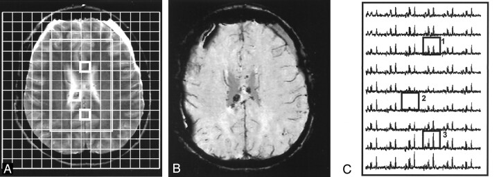
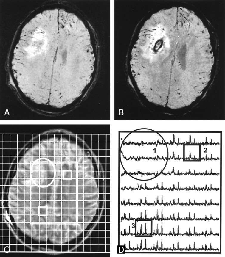
Methods: Forty children with TBI underwent transverse proton MRSI through the level of the corpus callosum within 1-16 days after injury. T2-weighted, fluid-attenuated inversion recovery, and susceptibility-weighted MR imaging was used to identify voxels as normal-appearing or as nonhemorrhagic or hemorrhagic injury. Neurologic outcome was evaluated at 6-12 months after injury. Metabolite ratios for total (all voxels), normal-appearing, and hemorrhagic brain were compared and used in a logistic regression model to predict long-term outcome. Total and regional metabolite ratios were compared with control data.
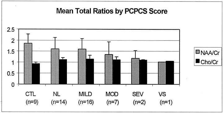
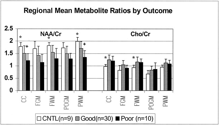
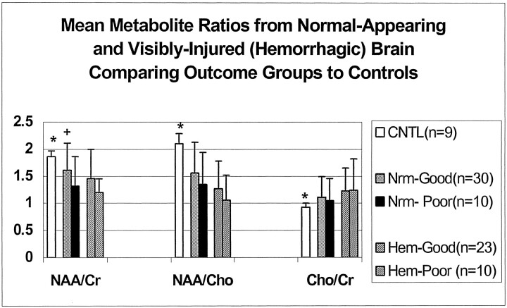
Results: A significant decrease in N-acetylaspartate (NAA)/creatine (Cr) and increase in choline (Cho)/Cr (evidence of DAI) was observed in normal-appearing (P < .05) and visibly injured (hemorrhagic) brain (P < .001) compared with controls. In normal-appearing brain NAA/Cr decreased more in patients with poor outcomes (1.32 +/- 0.54) than in those with good outcomes (1.61 +/- 0.50, P = .01) or control subjects (1.86 +/- 0.1, P = .00). In visibly injured brains, ratios were similarly altered in all patients. In predicting outcomes, ratios from normal-appearing and visibly-injured brain were 85% and 67% accurate, respectively.
Conclusion: MRSI can depict injury in brain that appears normal on imaging and is useful for predicting long-term outcomes.
Figures

Similar articles
-
Diffuse axonal injury in mild traumatic brain injury: a 3D multivoxel proton MR spectroscopy study.J Neurol. 2013 Jan;260(1):242-52. doi: 10.1007/s00415-012-6626-z. Epub 2012 Aug 12. J Neurol. 2013. PMID: 22886061 Free PMC article.
-
Prospective longitudinal proton magnetic resonance spectroscopic imaging in adult traumatic brain injury.J Magn Reson Imaging. 2006 Jul;24(1):33-40. doi: 10.1002/jmri.20607. J Magn Reson Imaging. 2006. PMID: 16755529
-
Assessment of mitochondrial impairment in traumatic brain injury using high-resolution proton magnetic resonance spectroscopy.J Neurosurg. 2008 Jan;108(1):42-52. doi: 10.3171/JNS/2008/108/01/0042. J Neurosurg. 2008. PMID: 18173309 Clinical Trial.
-
Diffuse axonal injury after traumatic brain injury is a prognostic factor for functional outcome: a systematic review and meta-analysis.Brain Inj. 2018;32(4):395-402. doi: 10.1080/02699052.2018.1429018. Epub 2018 Jan 30. Brain Inj. 2018. PMID: 29381396
-
Susceptibility-weighted imaging and proton magnetic resonance spectroscopy in assessment of outcome after pediatric traumatic brain injury.Arch Phys Med Rehabil. 2006 Dec;87(12 Suppl 2):S50-8. doi: 10.1016/j.apmr.2006.07.275. Arch Phys Med Rehabil. 2006. PMID: 17140880 Review.
Cited by
-
MRS Reveals Chronic Inflammation in T2w MRI-Negative Perilesional Cortex - A 6-Months Multimodal Imaging Follow-Up Study.Front Neurosci. 2019 Aug 16;13:863. doi: 10.3389/fnins.2019.00863. eCollection 2019. Front Neurosci. 2019. PMID: 31474824 Free PMC article.
-
Longitudinal Neuroimaging in Pediatric Traumatic Brain Injury: Current State and Consideration of Factors That Influence Recovery.Front Neurol. 2019 Dec 13;10:1296. doi: 10.3389/fneur.2019.01296. eCollection 2019. Front Neurol. 2019. PMID: 31920920 Free PMC article. Review.
-
Comparison of inter subject variability and reproducibility of whole brain proton spectroscopy.PLoS One. 2014 Dec 17;9(12):e115304. doi: 10.1371/journal.pone.0115304. eCollection 2014. PLoS One. 2014. PMID: 25517503 Free PMC article.
-
Longitudinal Metabolite Changes after Traumatic Brain Injury: A Prospective Pediatric Magnetic Resonance Spectroscopic Imaging Study.J Neurotrauma. 2019 Apr 15;36(8):1352-1360. doi: 10.1089/neu.2018.5919. Epub 2018 Dec 20. J Neurotrauma. 2019. PMID: 30351247 Free PMC article.
-
Cerebral microhemorrhages in a collegiate football player: clinical implications in the management of sports concussion.Sports Health. 2010 Sep;2(5):391-4. doi: 10.1177/1941738110374628. Sports Health. 2010. PMID: 23015965 Free PMC article.
References
-
- Adams JH, Graham DI, Murray LS, Scott G. Diffuse axonal injury due to nonmissile head injury in humans: an analysis of 45 cases. Ann Neurol 1982;12:557–563 - PubMed
-
- Adams JH, Doyle D, Ford I, Gennarelli TA, Graham DI, McLellan DR. Diffuse axonal injury in head injury: definition, diagnosis and grading. Histopathology 1989;15:49–59 - PubMed
-
- Graham DI, McIntosh TK, Maxwell WL, et al. Recent advances in neurotrauma. J Neuropathol Ex Neuro 2000;59:641–651 - PubMed
-
- Meythaler JM, Peduzzi JD, Eleftheriou E, Novack TA. Current concepts: diffuse axonal injury-associated traumatic brain injury. Arch Phys Med Rehabil 2000;82:1461–1471 - PubMed
-
- Gorrie C, Oakes S, Duflou J, Blumbergs P, Waite PME. Axonal injury in children after motor vehicle crashes: Extent, distribution, and size of axonal swellings using β-APP immunohistochemistry. J Neurotrauma 2002;19:1171–1182 - PubMed
MeSH terms
LinkOut - more resources
Full Text Sources