Exercise-induced gene expression changes in the rat spinal cord
- PMID: 15892452
- PMCID: PMC6009109
- DOI: 10.3727/000000005783992115
Exercise-induced gene expression changes in the rat spinal cord
Abstract
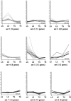
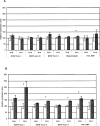
There is growing evidence that exercise benefits recovery of neuromuscular function from spinal cord injury (SCI). However, the effect of exercise on gene expression in the spinal cord is poorly understood. We used oligonucleotide microarrays to compare thoracic and lumbar regions of spinal cord of either exercising (voluntary wheel running for 21 days) or sedentary rats. The expression data were filtered using statistical tests for significance, and K-means clustering was then used to segregate lists of significantly changed genes into sets based upon expression patterns across all experimental groups. Levels of brain-derived neurotrophic factor (BDNF) protein were also measured after voluntary exercise, across different regions of the spinal cord. BDNF mRNA increased with voluntary exercise, as has been previously shown for other forms of exercise, contributed to by increases in both exon I and exon III. The exercise-induced gene expression changes identified by microarray analysis are consistent with increases in pathways promoting neuronal health, signaling, remodeling, cellular transport, and development of oligodendrocytes. Taken together these data suggest cellular pathways through which exercise may promote recovery in the SCI population.
Figures


References
-
- Abe Y.; Nakamura H.; Yoshino O.; Oya T.; Kimura T. Decreased neural damage after spinal cord injury in tPA-deficient mice. J Neurotrauma. 20:43–57; 2003. - PubMed
-
- Adlard P. A.; Cotman C. W. Voluntary exercise protects against stress-induced decreases in brain-derived neurotrophic factor protein expression. Neuroscience 124:985–992; 2004. - PubMed
-
- Adlard P. A.; Perreau V. M.; Engesser-Cesar C.; Cotman C. W. The timecourse of induction of brain-derived neurotrophic factor mRNA and protein in the rat hippocampus following voluntary exercise. Neurosci Lett. 363:43–48; 2004. - PubMed
-
- Ankeny D. P.; McTigue D. M.; Guan Z.; Yan Q.; Kinstler O.; Stokes B. T.; Jakeman L. B. Pegylated brain-derived neurotrophic factor shows improved distribution into the spinal cord and stimulates locomotor activity and morphological changes after injury. Exp. Neurol. 170:85–100; 2001. - PubMed
-
- Baldi P.; Long A. D. A Bayesian framework for the analysis of microarray expression data: Regularized t-test and statistical inferences of gene changes. Bioinformatics 17:509–519; 2001. - PubMed
Publication types
MeSH terms
Substances
Grants and funding
LinkOut - more resources
Full Text Sources
