The Open Microscopy Environment (OME) Data Model and XML file: open tools for informatics and quantitative analysis in biological imaging
- PMID: 15892875
- PMCID: PMC1175959
- DOI: 10.1186/gb-2005-6-5-r47
The Open Microscopy Environment (OME) Data Model and XML file: open tools for informatics and quantitative analysis in biological imaging
Abstract
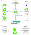
The Open Microscopy Environment (OME) defines a data model and a software implementation to serve as an informatics framework for imaging in biological microscopy experiments, including representation of acquisition parameters, annotations and image analysis results. OME is designed to support high-content cell-based screening as well as traditional image analysis applications. The OME Data Model, expressed in Extensible Markup Language (XML) and realized in a traditional database, is both extensible and self-describing, allowing it to meet emerging imaging and analysis needs.
Figures


References
-
- Phair RD, Misteli T. Kinetic modelling approaches to in vivo imaging. Nat Rev Mol Cell Biol. 2001;2:898–907. - PubMed
-
- Lippincott-Schwartz J, Snapp E, Kenworthy A. Studying protein dynamics in living cells. Nat Rev Mol Cell Biol. 2001;2:444–456. - PubMed
-
- Wouters FS, Verveer PJ, Bastiaens PI. Imaging biochemistry inside cells. Trends Cell Biol. 2001;11:203–211. - PubMed
-
- Ponti A, Machacek M, Gupton SL, Waterman-Storer CM, Danuser G. Two distinct actin networks drive the protrusion of migrating cells. Science. 2004;305:1782–1786. - PubMed
Publication types
MeSH terms
Grants and funding
LinkOut - more resources
Full Text Sources
Other Literature Sources

