. 2005 May 13:5:31.
doi: 10.1186/1471-2148-5-31.

The integrins of the urochordate Ciona intestinalis provide novel insights into the molecular evolution of the vertebrate integrin family

Affiliations

The integrins of the urochordate Ciona intestinalis provide novel insights into the molecular evolution of the vertebrate integrin family

Richard Ewan et al. BMC Evol Biol. .

Abstract

Background: Integrins are a functionally significant family of metazoan cell surface adhesion receptors. The receptors are dimers composed of an alpha and a beta chain. Vertebrate genomes encode an expanded set of integrin alpha and beta chains in comparison with protostomes such as drosophila or the nematode worm. The publication of the genome of a basal chordate, Ciona intestinalis, provides a unique opportunity to gain further insight into how and when the expanded integrin supergene family found in vertebrates evolved.

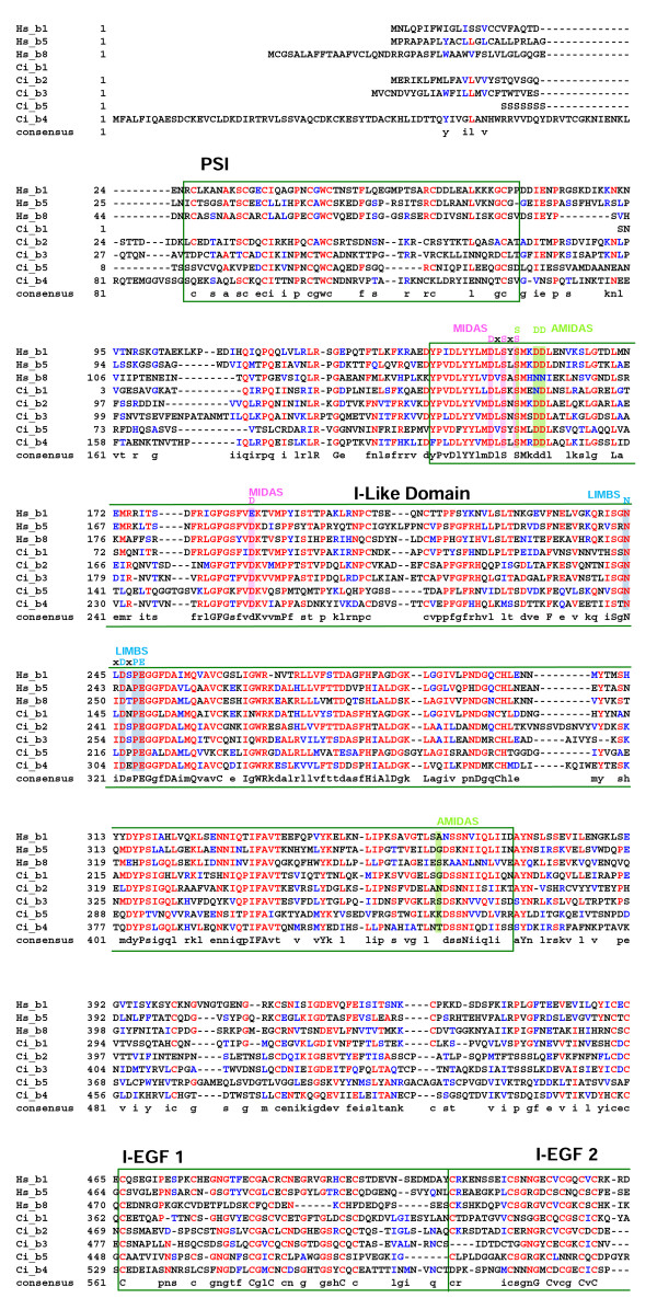
Results: The Ciona genome encodes eleven alpha and five beta chain genes that are highly homologous to their vertebrate homologues. Eight of the alpha chains contain an A-domain that lacks the short alpha helical region present in the collagen-binding vertebrate alpha chains. Phylogenetic analyses indicate the eight A-domain containing alpha chains cluster to form an ascidian-specific clade that is related to but, distinct from, the vertebrate A-domain clade. Two Ciona alpha chains cluster in laminin-binding clade and the remaining chain clusters in the clade that binds the RGD tripeptide sequence. Of the five Ciona beta chains, three form an ascidian-specific clade, one clusters in the vertebrate beta1 clade and the remaining Ciona chain is the orthologue of the vertebrate beta4 chain.

Conclusion: The Ciona repertoire of integrin genes provides new insight into the basic set of these receptors available at the beginning of vertebrate evolution. The ascidian and vertebrate alpha chain A-domain clades originated from a common precursor but radiated separately in each lineage. It would appear that the acquisition of collagen binding capabilities occurred in the chordate lineage after the divergence of ascidians.


Figures

Figure 1
Figure 1
Comparison of sequences recovered directly from the JGI C. intestinalis database before (A – Recovered) and after (B – Refined) sequence refinement. The annotated cartoons represent the domain structures of generic α & β integrin chains. Each subsequent row represents: (A – Recovered) the domain structure encoded by a sequence as retrieved directly from the database together with its assigned database accession number; and (B – Refined) the refined version of that gene after detailed analysis of the genomic sequence as described in the methods together with the name assigned during these analyses (e.g. Ci_α1). Refined sequences are presented as alignments in Fig. 3-7 and are also included as amino acid residue sequence files (see additional file 1).
Figure 2
Figure 2
The genomic locations and orientations of recovered and refined α integrin genes present on scaffolds 21 and 91. The genomic locations and orientations of the four very closely related A-domain containing α integrin genes identified after sequence refinement (Ci-α5–8, coloured red) together with the original five JGI-predicted gene fragments (blue) are indicated.
Figure 3
Figure 3
Alignment of the refined Ciona α chain sequences with representative human orthologues (residues 1-391 based on human α1 integrin chain). Protein domains and conserved motifs are annotated. Levels of sequence conservation are indicated (>50% identical, red; conservative substitutions, blue). MIDAS and α C-helix within the inserted A-domain are highlighted, as are the β-propeller domains 1–3.
Figure 4
Figure 4
Alignment of the refined Ciona α chain sequences with representative human orthologues (residues 392-796 based on human α1 integrin chain). Protein domains and conserved motifs are annotated. Levels of sequence conservation are indicated (>50% identical, red; conservative substitutions, blue). Ca2+-binding motifs in β-propeller repeats 5–7 are highlighted.
Figure 5
Figure 5
Alignment of the refined Ciona α chain sequences with representative human orthologues (residues 797 to C-terminus based on human α1 integrin chain). Protein domains and conserved motifs are annotated. Levels of sequence conservation are indicated (>50% identical, red; conservative substitutions, blue). Transmembrane domain (TM) and cytoplasmic interaction motif are indicated.
Figure 6
Figure 6
Alignment of the refined Ciona β chain sequences with representative human orthologues (residues 1-542 based on the human β1 integrin chain). Protein domains and conserved motifs are annotated. Levels of sequence conservation are indicated (>50% identical, red; conservative substitutions, blue). Adjacent to MIDAS (AMIDAS), ligand associated metal binding site (LIMBS) and MIDAS cation binding sites, and interaction motifs are highlighted as are the plexin/semaphorin/integrin (PSI), β-A domain (I-like) and epidermal growth factor (EGF) domains 1–2.
Figure 7
Figure 7
Alignment of the refined Ciona β chain sequences with representative human orthologues (residues 543 to the C-terminus based on the human β1 integrin chain). Protein domains and conserved motifs are annotated. Levels of sequence conservation are indicated (>50% identical, red; conservative substitutions, blue). EGF domains 2–4, transmembrane (TM) domain, interaction and phosphotyrosine binding (PTB) motifs are indicated.
Figure 8
Figure 8
Phylogenetic relationship of Ciona α integrin chains with representative protostome and vertebrate orthologues. Maximum Likelihood tree is shown with supporting Neighbor Joining bootstrap replicates (red) and Bayesian clade credibility values (green). Horizontal scale is amino acid replacements per site.
Figure 9
Figure 9
Phylogenetic relationship of Ciona β integrin chains with representative deuterostome orthologues. Maximum Likelihood tree is shown with supporting Neighbor Joining bootstrap replicates (red) and Bayesian clade credibility values (green). Horizontal scale is amino acid replacements per site.
Figure 10
Figure 10
Prediction of dimerisation patterns for novel integrin chains based on a combination of known interactions and phylogeny. A. Schematic phylogenies of α and β chains with established heterodimer pairing indicated by adjoining solid colored lines. B. Heterodimer pairings predicted (dashed lines) on the basis of the data presented in A. The color coding in B related to the known parings in A used to make the prediction.

References

    1. Mould AP, Humphries MJ. Regulation of integrin function through conformational complexity: not a simple knee-jerk interaction? Curr Opin Cell Biol. 2004;16:544–551. doi: 10.1016/j.ceb.2004.07.003. - DOI - PubMed
    1. Hynes RO. Integrins: Bidirectional, Allosteric Signaling Machines. Cell. 2002;110:673–687. doi: 10.1016/S0092-8674(02)00971-6. - DOI - PubMed
    1. Danen EHJ, Sonnenberg A. Integrins in regulation of tissue development and function. J Path. 2003;200:471–480. doi: 10.1002/path.1416. - DOI - PubMed
    1. Harris ES, McIntyre TM, Prescott SM, Zimmerman GA. The Leukocyte Integrins. J Biol Chem. 2000;275:23409–23412. doi: 10.1074/jbc.R000004200. - DOI - PubMed
    1. Schwartz MA, Assoian RK. Integrins and cell proliferation: regulation of cyclin-dependent kinases via cytoplasmic-signalling pathways. J Cell Sci. 2001;114:2553–2560. - PubMed

Publication types

LinkOut - more resources