Review
. 2005 Jul;96(1):9-21.
doi: 10.1093/aob/mci155. Epub 2005 May 13.

Cellulose biosynthesis: current views and evolving concepts

Affiliations
Review

Cellulose biosynthesis: current views and evolving concepts

Inder M Saxena et al. Ann Bot. 2005 Jul.

Abstract

Aims: To outline the current state of knowledge and discuss the evolution of various viewpoints put forth to explain the mechanism of cellulose biosynthesis. *

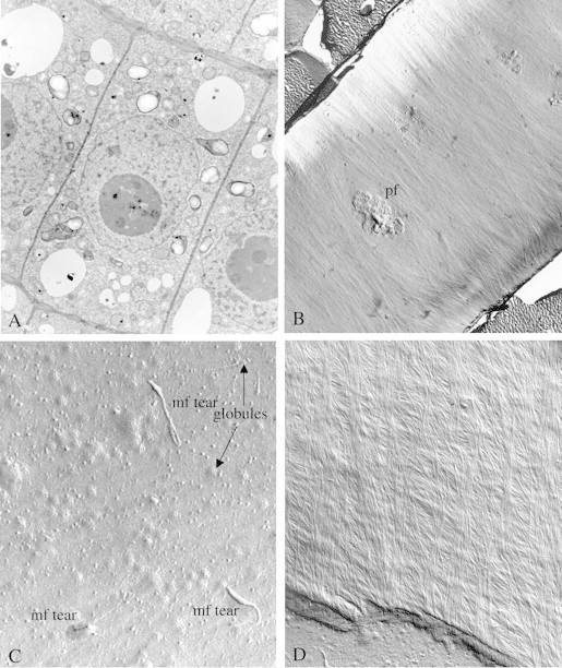
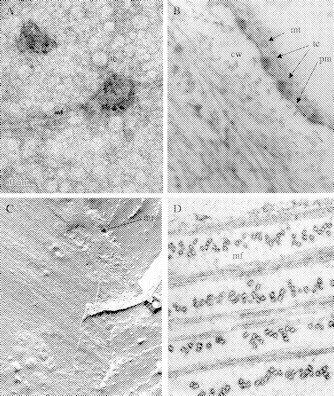
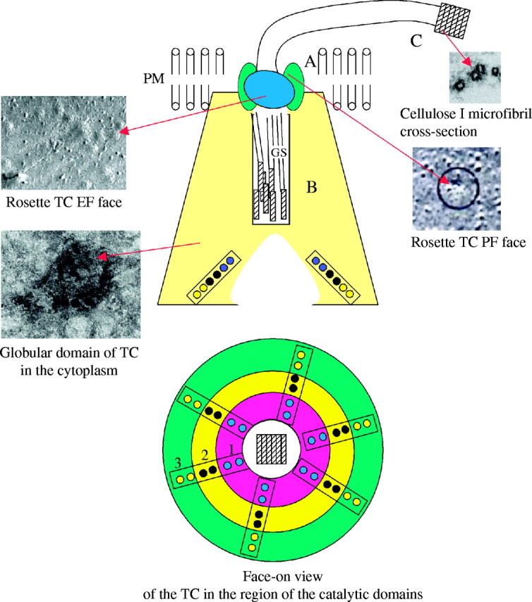
Scope: Understanding the mechanism of cellulose biosynthesis is one of the major challenges in plant biology. The simplicity in the chemical structure of cellulose belies the complexities that are associated with the synthesis and assembly of this polysaccharide. Assembly of cellulose microfibrils in most organisms is visualized as a multi-step process involving a number of proteins with the key protein being the cellulose synthase catalytic sub-unit. Although genes encoding this protein have been identified in almost all cellulose synthesizing organisms, it has been a challenge in general, and more specifically in vascular plants, to demonstrate cellulose synthase activity in vitro. The assembly of glucan chains into cellulose microfibrils of specific dimensions, viewed as a spontaneous process, necessitates the assembly of synthesizing sites unique to most groups of organisms. The steps of polymerization (requiring the specific arrangement and activity of the cellulose synthase catalytic sub-units) and crystallization (directed self-assembly of glucan chains) are certainly interlinked in the formation of cellulose microfibrils. Mutants affected in cellulose biosynthesis have been identified in vascular plants. Studies on these mutants and herbicide-treated plants suggest an interesting link between the steps of polymerization and crystallization during cellulose biosynthesis. *

Conclusions: With the identification of a large number of genes encoding cellulose synthases and cellulose synthase-like proteins in vascular plants and the supposed role of a number of other proteins in cellulose biosynthesis, a complete understanding of this process will necessitate a wider variety of research tools and approaches than was thought to be required a few years back.


Figures

F<sc>ig</sc>. 1.
Fig. 1.
(A) Ultrathin section of recently divided cells just below the meristem of a Zea mays root tip. Note the recently synthesized transverse walls (thinner). The elongation axis will be perpendicular to this direction. (Unpublished micrograph, courtesy of Susette Mueller and R. Malcolm Brown, Jr.) (B) Freeze fracture showing the E fracture face (EF) of a large area of an elongating cell in the root of Zea mays. The direction of microfibril impressions and, hence, the direction of the orientation of the microfibrils themselves, is perpendicular to the axis of elongation. Note also a prominent pit field (pf) in the centre of the micrograph. Microfibril synthesis around this pit field gives clues that suggest a membrane flow mechanism in the plane of the fluid membrane may underlie and direct cellulose microfibril synthesis (see Mueller and Brown, 1982a, b). Evidence to support this hypothesis is based on the direction of microfibrillar tears through the plasma membrane where the terminal globules and direction of synthesis is revealed (see C). In addition, parallel cortical microtubules provide the general ‘channels’ for the membrane flow. Actin microfilaments are found perpendicular to the cortical microtubules and may be the source of motion to propel the directional motions of the fluid membrane. (Unpublished micrograph, courtesy of Susette Mueller and R. Malcolm Brown, Jr.) (C) E fracture face of the plasma membrane of an actively elongating cell in the root of Zea mays showing three prominent tears of microfibrils back through the outer leaflet of the plasma membrane (mf tear). Note that the ‘rip’ terminates at a hole where the microfibril is associated with the rosette TC. In this fracture face, only the globular regions of the tips are shown associated with the TCs (globules). Many other TCs which have not been torn through the plasma membrane are revealed, some in clusters. (Unpublished micrograph, courtesy of Susette Mueller and R. Malcolm Brown, Jr.) (D) Freeze fracture through the innermost layer of a growth wall from an elongating cell in the root of Zea mays. Note the change in pitch of the transverse walls, suggesting that during elongation, the general pitch of the direction of microfibril synthesis is gradually changing from transverse to longitudinal. (Unpublished micrograph, courtesy of Susette Mueller and R. Malcolm Brown, Jr.)
F<sc>ig</sc>. 2.
Fig. 2.
(A) Negative staining of immunity-purified cellulose synthase from Gossypium hirsutum showing synthesis of cellulose I microfibrils in vitro. The identity of the isolated components of the rosette TC is demonstrated by immunolabelling using antibodies to CesA that are coupled with colloidal gold. The TC complex (tc) attached to a cellulose I microfibril is labelled with the antibody. When cellulose synthases are isolated using specific detergents and purified by immunoaffinity methods, they remain sufficiently intact to synthesize microfibrils (mf) as they would in vivo. This unpublished micrograph, courtesy of Walairat Laosinchai and R. Malcolm Brown, Jr, shows that the TC structure at the ‘business end’ is very different from the classical view of a rosette with a six-fold symmetry. (B) Ultrathin section through the plasma membrane of Boergesenia forbesii which has characteristic linear TCs, each with three rows of TC sub-units (see Kudlicka et al., 1987). In thin sections, these linear TCs can be observed in cross-section (tc), revealing structures never revealed by freeze fracture. In this case, a very large cytoplasmic component is imaged just beneath the plasma membrane (pm), and this proves that the typical TC structures revealed by freeze fractures show only ‘the tip of the iceberg’. These observations are consistent with the isolated functional TCs from Gossypium hirsutum (A) and form the basis for the revised model of TC structure/function (see Fig. 3). Note a single cortical microtubule (mt) adjacent to the plasma membrane and the cell wall (cw). (Unpublished micrograph, courtesy of Krystyna Kudlicka and R. Malcolm Brown, Jr.) (C) A multiple fracture through the cytoplasm and inner leaflet of the plasma membrane of an expanding cell in the root tip of Zea mays. This very unusual micrograph reveals the longitudinal fractures through cortical microtubules (mt) which parallel the underlying innermost layer of active microfibril synthesis. (Unpublished micrograph, courtesy of Susette Mueller and R. Malcolm Brown, Jr.) (D) Ultrathin section through the cell wall of the alga, Glaucocystis nostocherinum revealing the ordered arrangement of giant microfibrils (mf) synthesized by linear TCs. The alga synthesizes nearly pure cellulose Iα (Nishiyama et al., 2003). The microfibrils are synthesized in a complex helical pattern over the cell surface to reveal a precise rectangular shape. The microfibrils are coated with non-cellulose materials which stain well with a tannic acid post stain. These microfibrils are proposed to have more than 500 glucan chains per microfibril. While not identical to vascular plant cell walls, the Glaucocystis cell wall is perhaps one of the most beautiful examples to demonstrate the relationship between microfibril deposition and orientation to produce an ellipsoidal single cell. (Unpublished micrograph, courtesy of J. H. Martin Willison and R. Malcolm Brown, Jr.)
F<sc>ig</sc>. 3.
Fig. 3.
A revised model for the structure and function of the rosette TC in cellulose I microfibril biosynthesis. The 25-nm rosette portion of the TC (A) is shown in green where the six sub-units are largely localized to the innermost leaflet of the plasma membrane. The cytoplasmic portion of the TC is shown in yellow (B) and it contains the globular region of the catalytic sub-units. In this model, two identical sub-units of at least three different gene products form homodimers, all of which are required for cellulose I biosynthesis. Interestingly, the linear rows, each comprised of the three different cellulose synthases, are positioned such that the glucan chains produced by each sub-unit can rapidly associate by van der Waals interactions to produce the first stage of the crystalline cellulose product, namely a glucan chain sheet. Six separate glucan chain sheets are directed into the exit channel of the TC complex (B) where they pass through the rosette aperture and are then H-bonded into the crystalline cellulose I microfibril (C) that passes through this region to the surface of the cell. The face-on view of the cytoplasmic domain shows three different cellulose synthases, indicated as 1, 2 and 3, that are assembled as homodimers and organized in a linear row.

References

    1. Akashi T, Shibaoka. 1991. Involvement of transmembrane proteins in the association of cortical microtubules with the plasma membrane in tobacco BY-2 cells. Journal of Cell Science 98: 169–174.
    1. Albersheim P, Darvill A, Roberts K, Staehelin LA, Varner JE. 1997. Do the structures of cell wall polysaccharides define their mode of synthesis? Plant Physiology 113: 1–3. - PMC - PubMed
    1. Aloni Y, Delmer DP, Benziman M. 1982. Achievement of high rates of in vitro synthesis of 1,4-β-glucan: activation by cooperative interaction of the Acetobacter xylinum enzyme system with GTP, polyethylene glycol, and a protein factor. Proceedings of the National Academy of Sciences of the USA 79: 6448–6452. - PMC - PubMed
    1. Amikam D, Benziman M. 1989. Cyclic diguanylic acid and cellulose synthesis in Agrobacterium tumefaciens Journal of Bacteriology 177: 6649–6655. - PMC - PubMed
    1. Appenzeller L, Doblin M, Barreiro R, Wang H, Niu X, Kollipara K, et al. 2004. Cellulose synthesis in maize: isolation and expression analysis of the cellulose synthase (CesA) gene family. Cellulose 11: 287–299.

Publication types