. 2005 May 24;102(21):7517-22.
doi: 10.1073/pnas.0502495102. Epub 2005 May 13.

High-resolution x-ray crystal structures of the villin headpiece subdomain, an ultrafast folding protein

Affiliations

High-resolution x-ray crystal structures of the villin headpiece subdomain, an ultrafast folding protein

Thang K Chiu et al. Proc Natl Acad Sci U S A. .

Abstract

The 35-residue subdomain of the villin headpiece (HP35) is a small ultrafast folding protein that is being intensely studied by experiments, theory, and simulations. We have solved the x-ray structures of HP35 and its fastest folding mutant [K24 norleucine (nL)] to atomic resolution and compared their experimentally measured folding kinetics by using laser temperature jump. The structures, which are in different space groups, are almost identical to each other but differ significantly from previously solved NMR structures. Hence, the differences between the x-ray and NMR structures are probably not caused by lattice contacts or crystal/solution differences, but reflect the higher accuracy of the x-ray structures. The x-ray structures reveal important details of packing of the hydrophobic core and some additional features, such as cross-helical H bonds. Comparison of the x-ray structures indicates that the nL substitution produces only local perturbations. Consequently, the finding that the small stabilization by the mutation is completely reflected in an increased folding rate suggests that this region of the protein is as structured in the transition state as in the folded structure. It is therefore a target for engineering to increase the folding rate of the subdomain from approximately 0.5 micros(-1) for the nL mutant to the estimated theoretical speed limit of approximately 3 micros(-1).


Figures

Fig. 1.
Fig. 1.
Structure of WT (pH 6.7). (a and b) Front (a) and back (b) views. Uncharged, acidic, and basic residues are shown in black, red, and blue, respectively. Lighter shades of the same color show alternate conformations. Dashed lines depict H bonds <3.5 Å. (c) Detail of the hydrophobic core. Van der Waals contacts <4 Å are depicted as dashed lines, with each Phe and its neighbors in the same color. For clarity, only side-chain or main-chain atoms of some residues and hydrogens of the core Phes are shown. (d) A representative region of the final 2 FoFc map, contoured at 1.6 σ, showing density of D5–F6.
Fig. 2.
Fig. 2.
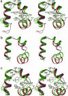
Comparison of x-ray and NMR structures. (a) 1VII (red) fitted to WT (pH 6.7) (green) by residues 1–34 with only selective side chains is shown. Dashed lines are H bonds as in Fig. 1. (b) The same structures are fitted by residues 23–33 to highlight their dramatic differences. (c) RMS fit as with a, but between WT (pH 6.7) (green) and 1QQV (red).
Fig. 3.
Fig. 3.
Thermodynamic and kinetic measurements. (a) Equilibrium thermal unfolding of WT (empty symbols) and nL (filled symbols) were monitored by the molar ellipticity at 222 nm and Trp fluorescence quantum yield (Φ, Inset). Black continuous lines are fits to the data using a two-state model {Keq = [U]/[F] = exp(–ΔH/R(1/T – 1/Tm)} (22). (Inset) Red squares show agreement of Φ values at equilibrium and after complete relaxation, indicating no slower kinetic phases. For nL, ΔH = –25 kcal/mol, Tm = 350 K, ΔS = 70 cal/(K mol), and ΔGfolding,300K = –3.6 kcal/mol (for WT, ΔGfolding,300K = –3.1 kcal/mol). (b) Φ for nL after a 10-ns T jump from 333 to 343 K (dashed blue line is equilibrium value at 343 K). The observed kinetics are biphasic, with the detail of the fast phase (Inset). (c) Two-state analysis of the slower relaxation phase (krel, black circles), which corresponds to overall unfolding/refolding kinetics (22). For nL, folding (blue circles): formula image; unfolding (red circles): formula image. (Inset) The same analysis for WT (22).
Fig. 4.
Fig. 4.
Packing of WT (pH 6.7) and structure of K24nL (pH 7.0). (a) A view down the crystallographic 2-fold axis in WT, with the Trp and Lys side chains in CPK. K24–D5 and W23–H27 interactions between symmetry-related molecules are depicted by dashed lines. (b) Front view of K24nL as in Fig. 1a. Note that the side chain of nL24 is compact here but extended in Fig. 1a. (c) Overlap of WT (green) and nL (red), but showing only the Phes, K29, and sulfate anions. (d) Same overlap as in c, but rendered as in Fig. 1b for all atoms.
Fig. 5.
Fig. 5.
Local perturbation by nL substitution. (a) A close-up view of residue 24 in WT (pH 6.7, blue) and K24nL (pH 7.0, yellow), showing the local perturbation of L28 by the K24nL mutation. (b) Plot of the difference in SASA for each residue between the two structures. Red and green bars correspond to residues that are involved or not involved in crystal contacts, respectively. (c) Differences in crystal contacts between the two structures are depicted in this molecular surface rendering, with residues involved in crystal contacts in red (observed in only one of two structures) or blue (observed in both structures) and residues not involved in any crystal contact in green.

Similar articles

Cited by

References

    1. Jones, C. M., Henry, E. R., Hu, Y., Chan, C. K., Luck, S. D., Bhuyan, A., Roder, H., Hofrichter, J. & Eaton, W. A. (1993) Proc. Natl. Acad. Sci. USA 90, 11860–11864. - PMC - PubMed
    1. Callender, R. H., Dyer, R. B., Gilmanshin, R. & Woodruff, W. H. (1998) Annu. Rev. Phys. Chem. 49, 173–202. - PubMed
    1. Gruebele, M. (1999) Annu. Rev. Phys. Chem. 50, 485–516. - PubMed
    1. Eaton, W. A., Munoz, V., Hagen, S. J., Jas, G. S., Lapidus, L. J., Henry, E. R. & Hofrichter, J. (2000) Annu. Rev. Biophys. Biomol. Struct. 29, 327–359. - PMC - PubMed
    1. Snow, C. D., Nguyen, H., Pande, V. S. & Gruebele, M. (2002) Nature 420, 102–106. - PubMed

Publication types

LinkOut - more resources