Comparative Study
. 2005 May 24;102(21):7565-70.
doi: 10.1073/pnas.0502031102. Epub 2005 May 13.

CDX1 is an important molecular mediator of Barrett's metaplasia

Affiliations
Comparative Study

CDX1 is an important molecular mediator of Barrett's metaplasia

N A C S Wong et al. Proc Natl Acad Sci U S A. .

Abstract

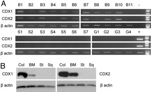
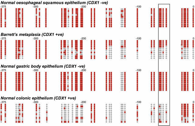
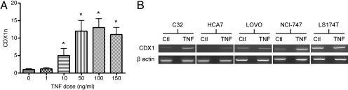
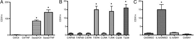
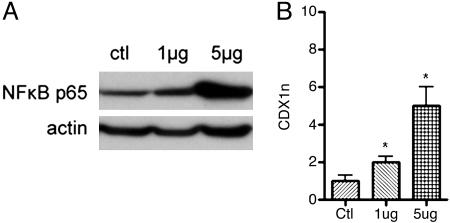
The molecular pathogenesis of Barrett's metaplasia (BM) of the esophagus is poorly understood. The change to an intestinal phenotype occurs on a background of esophagitis due to refluxing acid and bile. CDX1, an important regulator of normal intestinal development, was studied as a potential key molecule in the pathogenesis of BM. CDX1 mRNA and protein were universally expressed in all samples of BM tested but not in normal esophageal squamous or gastric body epithelia. This tissue-specific expression was attributable to the methylation status of the CDX1 promoter. Conjugated bile salts and the inflammatory cytokines TNF-alpha and IL-1beta were all found to increase CDX1 mRNA expression in vitro. These effects were primarily mediated by NF-kappaB signaling but only occurred when the CDX1 promoter was unmethylated or partially methylated. The data suggest that CDX1 is a key molecule linking etiological agents of BM to the development of an intestinal phenotype. Although the initial trigger for CDX1 promoter demethylation is not yet identified, it seems likely that demethylation of its promoter may be the key to the induction and maintenance of CDX1 expression and so of the BM phenotype.


Figures

Fig. 1.
Fig. 1.
CDX1 and CDX2 expression in gastrointestinal tissues. (A) CDX1, CDX2, and β-actin RT-PCRs performed on mRNA derived from normal esophageal squamous mucosal samples (S1–7), normal gastric mucosal samples (G1–4), and BM samples (B1–11). Positive (+) and water blank (–) controls and 100-bp ladders are all shown on the right side of the gels. (B) Examples of CDX1, CDX2, and β-actin Western blotting of lysates from normal colonic mucosa (Col), BM, normal gastric body mucosa (St), and normal esophageal squamous mucosa (Sq).
Fig. 2.
Fig. 2.
CDX1 promoter bisulfite sequencing of normal esophageal squamous epithelium, BM, normal gastric body epithelium, and normal colonic epithelium. For each tissue sample, relative location and methylation status of CDX1 promoter CpGs for 10 clones are shown. Each circle represents a CpG, and base positions relative to the CDX1 transcription start site are shown above each cluster of clones. Methylated CpGs are shaded. CpGs enclosed by the box (positions –53 to –65) represent those suggested to be crucial for transcriptional control (16).
Fig. 3.
Fig. 3.
TNF-α treatment of CRC cell lines. (A) CDX1 real-time PCR performed on HT29 cells treated with different doses of TNF-α for 5 h. The y axis (CDX1n) represents the amount of CDX1 normalized to GAPDH and relative to a calibrator (0 ng/ml sample). The error bars represent the calculated standard error of CDX1n for each sample. * denotes a significantly (at the 95% level) higher CDX1n of the sample compared with that of the calibrator. (B) CDX1 and β-actin RT-PCRs performed after TNF-α treatment of five CRC cell lines. Corresponding control-treated cell lines (Ctl) are also shown.
Fig. 4.
Fig. 4.
5aza2, signaling inhibitor, and cytokine treatments. (A) CDX1 real-time PCR performed on KYSE30 cells treated without 5aza2 or TNF-α, with 5aza2 or TNF-α, or with both. The calibrator used was the “control/control” treated sample. (B) CDX1 real-time PCR performed on four pairs of C32 cell cultures. Each pair was pretreated with an inhibitor of the NF-κB, ERK, c-Jun N-terminal kinase (JNK), or p38 MAPK signaling pathways. One of each pair of cultures was then treated with 50 ng/ml TNF-α (T) or control medium (C) for 5 h. The calibrator used for each pair was the control-treated sample. (C) CDX1 real-time PCR performed on C32 cells. The cells were pretreated with BAY11-7085 (BAY) or its carrier medium DMSO and then treated with 50 ng/ml IL-1β or control medium (Ctl). The calibrator used was the “control/DMSO” treated sample. The format of all three graphs is as for Fig. 3A.
Fig. 5.
Fig. 5.
Transfection of C32 cells with NF-κB p65. The cell line was transfected with 1 μg or 5 μg of NF-κB p65 expression vector or with empty vector as a treatment control (ctl). (A) Western blotting of nuclear lysates confirmed successful activation of NF-κB signaling as indicated by increased nuclear expression of NF-κB p65 protein (β-actin was used as a loading control). (B) CDX1 real-time PCR showed that C32 cells responded to the NF-κB p65 transfection-mediated expression with an increase in CDX1 mRNA expression. The format of the graph is as for Fig. 3A. The calibrator used was the “control” transfected sample.
Fig. 6.
Fig. 6.
Treatment of C32 cells with either acid (pH 3.5) or bile salts or both. Mock-treated C32 cells (ctl) served as a control. (A) Western blotting of nuclear lysates showed that bile salts alone, or acidified bile salts, activated NF-κB signaling. (B) The treatments were also associated with an increase in CDX1 mRNA expression as assessed by real-time PCR. The format of the graph is as for Fig. 3A. The calibrator used was the control treated sample.

Similar articles

Cited by

References

    1. Nandurkar, S. & Talley, N. J. (1999) Am. J. Gastroenterol. 94, 30–40. - PubMed
    1. Wild, C. P. & Hardie, L. J. (2003) Nat. Rev. Cancer 3, 676–684. - PubMed
    1. Freund, J. N., Domon-Dell, C., Kedinger, M. & Duluc, I. (1998) Biochem. Cell Biol. 76, 957–969. - PubMed
    1. Eda, A., Osawa, H., Yanaka, I., Satoh, K., Mutoh, H., Kihira, K. & Sugano, K. (2002) J. Gastroenterol. 37, 94–100. - PubMed
    1. Eda, A., Osawa, H., Satoh, K., Yanaka, I., Kihira, K., Ishino, Y., Mutoh, H. & Sugano, K. (2003) J. Gastroenterol. 38, 14–22. - PubMed

Publication types

MeSH terms

LinkOut - more resources