Comparative Study
. 2005 May 24;102(21):7766-71.
doi: 10.1073/pnas.0500113102. Epub 2005 May 13.

An ancestral oomycete locus contains late blight avirulence gene Avr3a, encoding a protein that is recognized in the host cytoplasm

Affiliations
Comparative Study

An ancestral oomycete locus contains late blight avirulence gene Avr3a, encoding a protein that is recognized in the host cytoplasm

Miles R Armstrong et al. Proc Natl Acad Sci U S A. .

Abstract

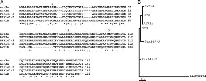
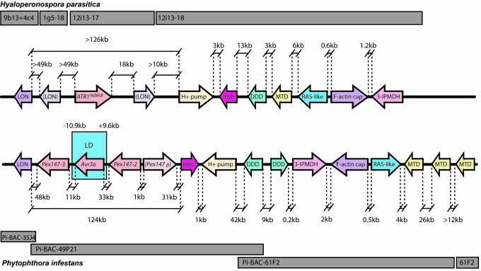
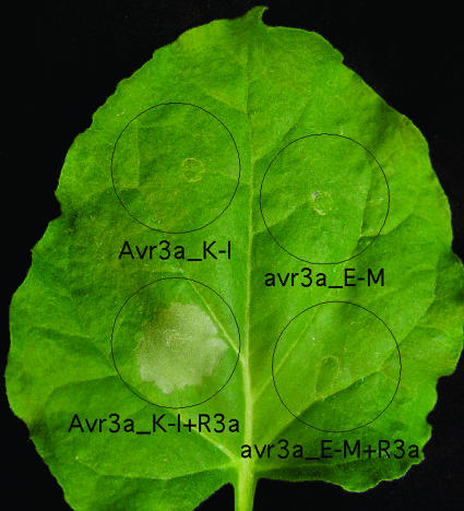
The oomycete Phytophthora infestans causes late blight, the potato disease that precipitated the Irish famines in 1846 and 1847. It represents a reemerging threat to potato production and is one of >70 species that are arguably the most devastating pathogens of dicotyledonous plants. Nevertheless, little is known about the molecular bases of pathogenicity in these algae-like organisms or of avirulence molecules that are perceived by host defenses. Disease resistance alleles, products of which recognize corresponding avirulence molecules in the pathogen, have been introgressed into the cultivated potato from a wild species, Solanum demissum, and R1 and R3a have been identified. We used association genetics to identify Avr3a and show that it encodes a protein that is recognized in the host cytoplasm, where it triggers R3a-dependent cell death. Avr3a resides in a region of the P. infestans genome that is colinear with the locus containing avirulence gene ATR1(NdWsB) in Hyaloperonospora parasitica, an oomycete pathogen of Arabidopsis. Remarkably, distances between conserved genes in these avirulence loci were often similar, despite intervening genomic variation. We suggest that Avr3a has undergone gene duplication and that an allele evading recognition by R3a arose under positive selection.


Figures

Fig. 1.
Fig. 1.
Protein alignments and phylogeny of AVR3a, avr3a, and PEX147-like paralogues. (A) Multiple alignment (using www.ebi.ac.uk/clustalw) of predicted proteins (AVR3a and avr3a) derived from the Avr3a (avirulent) and avr3a (virulent) alleles (initially termed Pex147) with those from the two paralogous P. infestans Pex147-2 and Pex147-3 sequences and the AVR1b protein from P. sojae. The N-terminal type II SP (first 21 aa) and the RXLR motif, found also in theH. parasitica ATR1NdWsB protein (32), are underlined. The locations of the three amino acid polymorphisms are indicated by numbers above the amino acids. (B) Maximum likelihood estimation of the phylogeny of Avr3a, avr3a, and Pex147-like paralogues using a nucleotide alignment based on the amino acid alignment in A. The tree was constructed by using dnaml from the phylip package, with the P. sojae Avr1b sequence (AAM20934) as an outgroup. Bootstrap values from 1,000 random resamplings of the data are indicated. The alignment supports the closer evolutionary relationship of the Avr3a allele, rather than the avr3a allele, to the flanking Pex147-2 and Pex147-3 paralogues.
Fig. 2.
Fig. 2.
Conservation of synteny between H. parasitica ATR1NdWsB (Upper) and P. infestans Avr3a (Lower) loci. Sequenced BACs are indicated as gray blocks. Arrows indicate positions and transcriptional orientation of CDSs encoding Lon protease (LON), partial LON sequences (bracketed), vacuolar proton ATPase (H+ PUMP), Myb transcription factor (myb), dimethyl dihydrodiol dehdrogenase (DDD), methylenetetrahydrofolate dehydrogenase (MTD), a RAB (RAS-like), F-actin capping protein (F-actin cap), and 3-isopropylmalate dehydrogenase (3-IPMDH). Distances (in kb) between sequences (not to scale) are indicated. Probes PCR-amplified (see Table 2 for primers) from each P. infestans sequence hybridized to the P. infestans BAC library (16) did not indicate colocalization of these sequences elsewhere in the genome (data not shown). The block of LD defined by primers 9.6-kb upstream and 10.9-kb downstream of Avr3a (initially termed Pex147) is indicated, as are the flanking Pex147-2 and Pex147-3 paralogues and a Pex147-like pseudogene (Pex147p).
Fig. 3.
Fig. 3.
Pex147 is up-regulated before and during potato infection. Real-time RT-PCR analysis of the expression of Pex147 (avr3a), using cDNA templates derived from P. infestans 88069 (virulent on R3a genotypes; Table 1) nonsporulating mycelium (M) grown in pea broth, sporangia (S), zoospores (Z), germinating cysts (C), germinating cysts forming appressoria (A), and infected susceptible cv. Bintje 12, 24, 33, 48, 56, and 72 h postinoculation.
Fig. 4.
Fig. 4.
Cobombardment demonstrates recognition of AVR3a by plants containing R3a. Truncated (E80 M103) avr3a and (K80 I103) Avr3a alleles were transiently coexpressed with gfp in potato genotypes containing R1 (a), R2 (b), R3b (c), R10 (d), and R3a (e), each compared with cv. Bintje (r), and in transgenic cv. Desiree expressing R3a compared with cv. Desiree (f). Typically, more GFP-expressing cells were observed on R gene-containing genotypes when gfp was bombarded alone than on adjacent Bintje leaves (ae), resulting in GFP fluorescence indexes >1 (where 1 is the average total GFP fluorescence on Bintje). In each histogram, the average GFP fluorescence index is presented after coexpression with Avr3a (light gray) or avr3a (dark gray) and expression of gfp alone (white).
Fig. 5.
Fig. 5.
Transient expression and coexpression of Avr3a and R3a in N. benthamiana. Leaves of N. benthamiana plants were infiltrated with A. tumefaciens carrying pGR106::Avr3a_K-I or pGR106::avr3a_E-M (expressing mature forms of the proteins lacking SPs) alone or mixed with an A. tumefaciens strain carrying pBINplus::R3a. For transient coexpression of Avr3a and R3a, the A. tumefaciens solutions were mixed in a 1:2 ratio before infiltration. Photographs of symptoms were taken 6 days postinfiltration. Circles indicate the infiltrated area on the leaf panels for each treatment. This is a representative leaf from multiple assays and experiments.

References

    1. Birch, P. R. J. & Whisson, S. C. (2001) Mol. Plant Pathol. 2, 257–263. - PubMed
    1. Schiermeier, Q. (2001) Nature 410, 1011. - PubMed
    1. Duncan, J. M. (1999) Microbiol. Today 26, 114–116.
    1. Kamoun, S. (2003) Eukaryot. Cell 2, 191–199. - PMC - PubMed
    1. Sogin, M. L. & Silberman, J. D. (1998) Int. J. Parasitol. 28, 11–20. - PubMed

Publication types

MeSH terms

Associated data

LinkOut - more resources