. 2005 Jun;17(6):1685-703.
doi: 10.1105/tpc.105.031393. Epub 2005 May 13.

Identification and functional analysis of in vivo phosphorylation sites of the Arabidopsis BRASSINOSTEROID-INSENSITIVE1 receptor kinase

Affiliations

Identification and functional analysis of in vivo phosphorylation sites of the Arabidopsis BRASSINOSTEROID-INSENSITIVE1 receptor kinase

Xiaofeng Wang et al. Plant Cell. 2005 Jun.

Abstract

Brassinosteroids (BRs) regulate multiple aspects of plant growth and development and require an active BRASSINOSTEROID-INSENSITIVE1 (BRI1) and BRI1-ASSOCIATED RECEPTOR KINASE1 (BAK1) for hormone perception and signal transduction. Many animal receptor kinases exhibit ligand-dependent oligomerization followed by autophosphorylation and activation of the intracellular kinase domain. To determine if early events in BR signaling share this mechanism, we used coimmunoprecipitation of epitope-tagged proteins to show that in vivo association of BRI1 and BAK1 was affected by endogenous and exogenous BR levels and that phosphorylation of both BRI1 and BAK1 on Thr residues was BR dependent. Immunoprecipitation of epitope-tagged BRI1 from Arabidopsis thaliana followed by liquid chromatography-tandem mass spectrometry (LC/MS/MS) identified S-838, S-858, T-872, and T-880 in the juxtamembrane region, T-982 in the kinase domain, and S-1168 in C-terminal region as in vivo phosphorylation sites of BRI1. MS analysis also strongly suggested that an additional two residues in the juxtamembrane region and three sites in the activation loop of kinase subdomain VII/VIII were phosphorylated in vivo. We also identified four specific BAK1 autophosphorylation sites in vitro using LC/MS/MS. Site-directed mutagenesis of identified and predicted BRI1 phosphorylation sites revealed that the highly conserved activation loop residue T-1049 and either S-1044 or T-1045 were essential for kinase function in vitro and normal BRI1 signaling in planta. Mutations in the juxtamembrane or C-terminal regions had only small observable effects on autophosphorylation and in planta signaling but dramatically affected phosphorylation of a peptide substrate in vitro. These findings are consistent with many aspects of the animal receptor kinase model in which ligand-dependent autophosphorylation of the activation loop generates a functional kinase, whereas phosphorylation of noncatalytic intracellular domains is required for recognition and/or phosphorylation of downstream substrates.


Figures
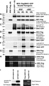
Figure 1.
Figure 1.
BL Dependence of Early Events in BR Signaling. Transgenic Arabidopsis plants expressing both BRI1-Flag and BAK1-GFP were grown as described in the text. Total membrane protein was purified from each sample, including an untreated, untransformed (wild-type) control, and two BL-treated lines expressing BRI-Flag or BAK1-GFP alone. Immunoprecipitation of solubilized membrane proteins was performed as described (Li et al., 2002), and equal amounts of protein were separated by SDS-PAGE followed by protein gel blot analysis as indicated. The entire experiment ([A] to [I]) was repeated three times with consistent results. Numbers to the left of each blot indicate migration of molecular mass markers (kD). (A) to (D) Approximately equal amounts of BRI1-Flag and BAK1-GFP were present in total membrane extracts and immunoprecipitated samples, indicating that BL or BRZ treatment did not affect the total level of these proteins in planta. (E) Association of BRI1 and BAK1 in vivo, as determined by coimmunoprecipitation, is BL dependent. (F) and (G) Phosphorylation of Thr residues in BRI1-FLAG and BAK1-GFP is BL dependent (note that the FLAG epitope tag has no Thr residues). (H) ProQ Diamond stain, which recognizes phosphorylated Ser and Thr residues, binds to BRI1-Flag immunoprecipitated from plants with reduced endogenous BL, suggesting that at least one Ser residue is constitutively phosphorylated in vivo. Sypro Ruby is a general protein stain that shows loading levels. (I) GST-BAK1-KD and Flag-BRI1-KD are recombinant cytoplasmic domains of BAK1 and BRI1 purified from bacteria and autophosphorylated in vitro. GST-mBAK1-KD and Flag-mBRI1-KD both have a K-to-E substitution in kinase subdomain II, resulting in complete inability to autophosphorylate in vitro. The phosphothreonine antibody recognizes only the phosphorylated form of BAK1. See Figure 6C for an identical result with BRI1. Similarly, ProQ Diamond recognizes only the phosphorylated form of BRI1.
Figure 2.
Figure 2.
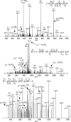
Ion Trap MS/MS Spectra Identifying BRI1 in Vivo Phosphorylation Sites. Eleven-day-old light-grown Arabidopsis plants expressing a full-length BRI1 gene with a C-terminal Flag epitope were treated with 0.1 μM BL, and BRI1-Flag was immunoprecipitated with anti-Flag antibody linked to agarose beads. The protein was PAGE purified and in-gel digested with trypsin, and the resulting peptides were extracted and analyzed by LC/MS/MS using a capillary LC system coupled directly to a LCQ Deca ion trap mass spectrometer. Each MS/MS spectrum is a collection of ions produced by collision-induced dissociation of the intact peptide. The fragmentation preferentially occurs at peptide bonds to generate N-terminal fragments (b ions) and C-terminal fragments (y ions) at specific m/z ratios and intensities that provide information regarding amino acid sequence and sites of modification as indicated by vertical lines between residues of the peptide sequence. The predominant b and y product ion peaks are labeled accordingly with the subscripts denoting their position in the identified peptide and the superscripts of + and ++ indicating singly and doubly protonated ions, respectively. Product ions eliciting neutral mass losses of H2O, NH3, and H3PO4 are also indicated. Identified phosphoseryl and phosphothreonyl resides are denoted as pS and pT, respectively. Met sulfoxide residues (oxidized Met) are labeled as M*. Analysis of y and b ion fragmentation patterns with SEQUEST showed that S-838, T-982, and S-1168 were phosphorylated in vivo.
Figure 3.
Figure 3.
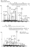
QTOF MS/MS Spectra Identifying BRI1 in Vivo Phosphorylation Sites. BRI1-Flag was immunoprecipitated from Arabidopsis plants as described in Figure 2. The protein was SDS-PAGE purified and in-gel digested with trypsin, and the resulting peptides were extracted and analyzed by LC/MS/MS analysis on a capillary LC system coupled directly to a Waters Q-ToF Ultima mass spectrometer (Milford, MA). The measured mass (D) of the singly protonated peptide and the corresponding mass measurement accuracy (ppm) are reported above each spectrum. The ion product spectrum corresponding to fragmentation of each singly charged peptide is shown. Prominent b and y ions are labeled as in Figure 2, including neutral loss of H3PO4 (98 D) from phosphorylated peptides as indicated. Analysis of y and b ion fragmentation patterns with Mascot showed that S-858, T-872, and T-880 were phosphorylated in vivo.
Figure 4.
Figure 4.
Summary of BRI1 Phosphorylation Sites. The juxtamembrane region and C-terminal region are italicized. The six confirmed sites of in vivo phosphorylation are outlined, and the five confirmed sites of in vitro phosphorylation (Oh et al., 2000) are underlined. Phosphorylated peptides with ambiguity in the specific residues are indicated in bold, with open triangles showing possible in vivo sites and closed triangles possible in vitro sites. The activation loop is marked with brackets and the peptide beginning with LMSAM is phosphorylated on three of the Ser or Thr residues in vivo. The peptide beginning with DTHLS is phosphorylated on three Ser or Thr residues in vitro. The peptide TANNTNWKLTGVK is phosphorylated on two of the three marked residues in vivo. All of the remaining peptides in bold are phosphorylated on only one of the indicated residues in vitro and on the specific residues outlined in vivo.
Figure 5.
Figure 5.
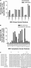
Conserved Ser and Thr Residues in the Arabidopsis RLK Family Corresponding to Possible BRI1 Phosphorylation Sites. (A) A PileUP protein alignment of the kinase domains of all 610 Arabidopsis RLKs (Shiu and Bleecker, 2001) was visually examined for the presence of Ser or Thr residues at specific sites in the alignment corresponding to the indicated BRI1 residues. A subset of 217 putative LRR RLKs was tabulated separately. (B) The intracellular domains of 121 Arabidopsis LRR RLKs containing an Arg immediately preceding the invariant Asp in the catalytic site of subdomain VI (RD kinases) were aligned with ClustalW, and the number of Ser and Thr residues occurring in the positions corresponding to the indicated BRI1 residues were tabulated. A similar analysis was performed with 92 non-RD Arabidopsis LRR RLKs. (C) Specific activation loop residues of RD LRR RLKs are highly conserved. PileUp was used to align sequences in 12 LRR RLKs (RD-type kinases) from the RD in subdomain VIb to the invariant E in subdomain VIII. Note the very high conservation of Ser or Thr residues at positions corresponding to BRI1 T-1039, S-1044, and T-1049.
Figure 6.
Figure 6.
Effect of Mutating Specific Ser and Thr Residues of the BRI1 Cytoplasmic Domain on Autophosphorylation and Substrate Phosphorylation in Vitro. (A) Autophosphorylation of recombinant Flag-BRI1-KD (wild type) and a range of mutants, including a kinase inactive mutant (mBRI1-KD) and constructs in which specific Ser and Thr residues were substituted with Ala by site-directed mutagenesis. Equal amounts of recombinant protein (as indicated by the stained gel in the top panel) were incubated with [γ32P]ATP and separated by SDS-PAGE, followed by autoradiography (bottom panel). The experiment was repeated three times with consistent results. (B) Phosphorylation of a synthetic peptide (BR13) containing the consensus sequence for optimum BRI1-KD substrate phosphorylation. Typically, a 40-μL reaction contained BRI1-KD or the indicated mutant protein (1.0 μg), 0.1 mM [γ-32P]ATP (500 cpm/pmol), and synthetic peptide (100 μg mL−1) in kinase buffer. Incorporation of 32P into the synthetic peptide was quantified by binding to P81 phosphocellulose paper. Reactions were performed in triplicate. Error bars indicate se. (C) Hyperautophosphorylation of Flag-BRI1(T-872-A) in E. coli. Recombinant proteins were purified from E. coli with anti-Flag M2 antibody attached to agarose beads, and 0.1 μg of recombinant protein was separated by SDS-PAGE. After transfer to polyvinylidene difluoride (PVDF) membranes, immunoblot analysis was performed with anti-Flag antibody to demonstrate equal protein transfer and antiphosphothreonine antibody to determine autophosphorylation levels. Note that the antibody was specific for the phosphorylated form of the protein (no signal with the kinase inactive mutant K-911-E, Flag-mBRI-KD) and that the high level of phosphosphorylation of the T-872-A mutation caused a mobility shift during SDS-PAGE.
Figure 7.
Figure 7.
Effect of Mutating Specific Ser and Thr Residues of the BRI1 Cytoplasmic Domain on the Kinetics of BR13 Peptide Substrate Phosphorylation in Vitro. Both Km and Vmax for BR13 were dramatically altered by substituting Ala for Ser or Thr at specific sites in the juxtamembrane or C-terminal regions of BRI1-KD. T-842-A and S/T-1179/80-A showed less catalytic activity, whereas T-872-A showed an ∼10-fold increase in Vmax and twofold reduction in Km, indicating a much higher enzyme efficiency compared with the wild type.
Figure 8.
Figure 8.
Effect of Mutating Specific Ser and Thr Residues of the BRI1 Cytoplasmic Domain on Rescue of the bri1-5 Mutant. Transgenic constructs contained 1699 bp of 5′ upstream BRI1 sequence (covering the BRI1 promoter and 5′ untranslated region), the entire coding region, and an in-frame C-terminal epitope tag (WT BRI1-Flag). A kinase-inactive version of the same construct was generated by a K-911-E mutation (mBRI1-Flag). Substitution of Ala for specific Thr or Ser residues within the wild-type construct are as indicated. Three independent transgenic lines for each construct are shown. All lines were grown under the same conditions and are the same age (32 d). Transgene expression was monitored by immunoblot analysis with anti-Flag antibody using equal amounts of protein from each plant. Cosuppression was examined by assaying native BRI1 RNA levels using competitive RT-PCR on equal amounts of total RNA isolated from each plant. The synthetic RNA competitor had the same sequence (except for a 53-nucleotide deletion in the center of the fragment) and used the same primer pair as endogenous BRI1. Lanes marked (−) had no reverse transcriptase and verified lack of DNA contamination.

References

    1. Adams, J.A. (2003). Activation loop phosphorylation and catalysis in protein kinases: Is there functional evidence for the autoinhibitor model? Biochemistry 42, 601–607. - PubMed
    1. Altmann, T. (1999). Molecular physiology of brassinosteroids revealed by the analysis of mutants. Planta 208, 1–11. - PubMed
    1. Asami, T., Min, Y.K., Nagata, N., Yamagishi, K., Takatsuto, S., Fujioka, S., Murofushi, N., Yamaguchi, I., and Yoshida, S. (2000). Characterization of brassinazole, a triazole-type brassinosteroid biosynthesis inhibitor. Plant Physiol. 123, 93–100. - PMC - PubMed
    1. Baxter, R.M., Secrist, J.P., Vaillancourt, R.R., and Kazlauskas, A. (1998). Full activation of the platelet-derived growth factor beta-receptor kinase involves multiple events. J. Biol. Chem. 273, 17050–17055. - PubMed
    1. Becker, D., Kemper, E., Schell, J., and Masterson, R. (1992). New plant binary vectors with selectable markers located proximal to the left T-DNA border. Plant Mol. Biol. 20, 1195–1197. - PubMed

Publication types

MeSH terms