. 2005 Jun;138(2):767-77.
doi: 10.1104/pp.104.055756. Epub 2005 May 13.

Arabidopsis thickvein mutation affects vein thickness and organ vascularization, and resides in a provascular cell-specific spermine synthase involved in vein definition and in polar auxin transport

Affiliations

Arabidopsis thickvein mutation affects vein thickness and organ vascularization, and resides in a provascular cell-specific spermine synthase involved in vein definition and in polar auxin transport

Nicole K Clay et al. Plant Physiol. 2005 Jun.

Abstract

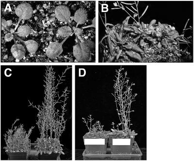
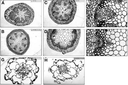
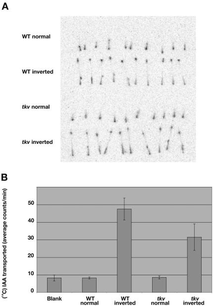
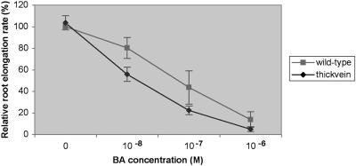
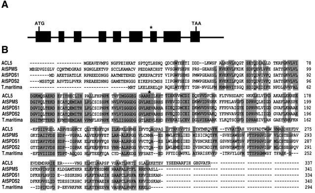
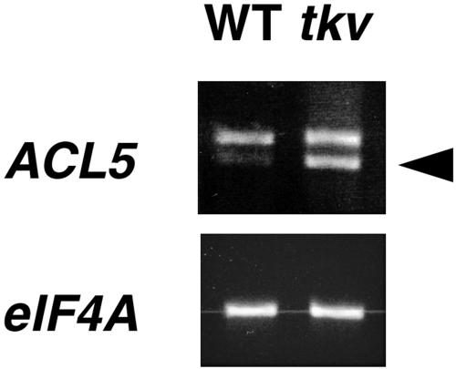
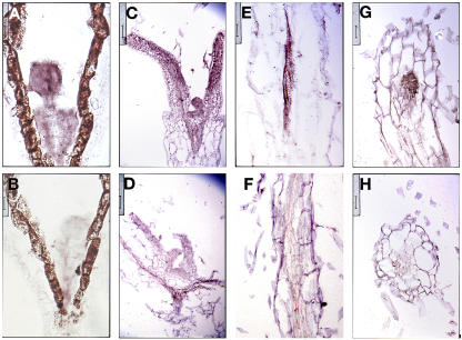
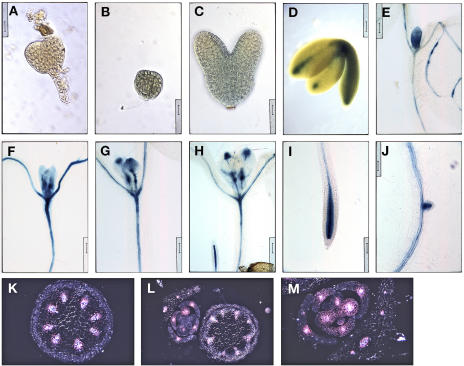
Polar auxin transport has been implicated in the induction of vascular tissue and in the definition of vein positions. Leaves treated with chemical inhibitors of polar auxin transport exhibited vascular phenotypes that include increased vein thickness and vascularization. We describe a recessive mutant, thickvein (tkv), which develops thicker veins in leaves and in inflorescence stems. The increased vein thickness is attributable to an increased number of vascular cells. Mutant plants have smaller leaves and shorter inflorescence stems, and this reduction in organ size and height is accompanied by an increase in organ vascularization, which appears to be attributable to an increase in the recruitment of cells into veins. Furthermore, although floral development is normal, auxin transport in the inflorescence stem is significantly reduced in the mutant, suggesting that the defect in auxin transport is responsible for the vascular phenotypes. In the primary root, the veins appear morphologically normal, but root growth in the tkv mutant is hypersensitive to exogenous cytokinin. The tkv mutation was found to reside in the ACL5 gene, which encodes a spermine synthase and whose expression is specific to provascular cells. We propose that ACL5/TKV is involved in vein definition (defining the boundaries between veins and nonvein regions) and in polar auxin transport, and that polyamines are involved in this process.


Figures

Figure 1.
Figure 1.
Increased vein thickness and vascularization in tkv leaves. A to H, Cleared leaves of wild type (A, C, and E), tkv (B, D, F, and G), and fully restored tkv containing wild-type copy of the ACL5 transgene (H) were viewed under dark-field illumination to visualize the venation pattern. A and B, First pair of juvenile leaves. C, D, G and H, Second pair of juvenile leaves. E and F, Adult leaves. All leaves are in same magnification, and leaves used for comparison are developmentally equivalent (have similar number of secondary veins that form prominent arches that begin and end at the midvein) and representative of numerous samples. I to O, Transverse sections through the midvein (I, J, and M–O) and lateral veins (K and L) of fully expanded juvenile leaves. I, K, and M, Wild type. J, L, N, and O, tkv. Arrowheads indicate the midvein, and arrow indicates a secondary vein. In wild type, aside from the midvein, higher-order veins are indistinguishable by vein thickness. Scale bars in I to L = 50 μm, and scale bars in M to O = 16 μm.
Figure 2.
Figure 2.
Shortened inflorescence stems of tkv mutant. A, Two-week-old wild-type (left) and tkv (right) plants. B, Top view of 4-week-old wild-type (left) and tkv (two on right) plants. C, Fully grown tkv (left) and wild-type (right) plants. D, Fully grown tkv plants (left) and tkv plants containing the wild-type ACL5 transgene (right).
Figure 3.
Figure 3.
Increased cambium in inflorescence stems of tkv mutant. A to F, Transverse sections through the apical end of the inflorescence stem (just below the youngest silique; A and B) and the basal end of the inflorescence stem (C, D, E, and F). A, C, E, tkv. B, D, F, Wild type. E and F are enlargements of a vascular bundle in C and D, respectively. Opaque cells inside of the phloem are cambial-like cells and possibly include xylem parenchyma cells. Arrowheads point to xylem, and arrows point to phloem. Scale bars in A, B, C, D, G, and H = 50 μm, and scale bars in E and F = 25 μm. G and H, Transverse sections through the primary root of week-old seedlings in the root elongation zone, where root hairs emerge. G, Wild type. H, tkv. Arrowheads point to xylem axes, and arrows point to phloem cells. Scale bars = 16 μm.
Figure 4.
Figure 4.
Reduced polar auxin transport in inflorescence stems of tkv mutant. A, The basipetal (inverted orientation) and acropetal (normal orientation) transport of 14C-IAA in excised inflorescence stem segments were visualized using the Fujix image analyzer. B, Average cpm of 14C-IAA transported to the distal end of the stem segments were calculated from 80 samples of each genotype from four separate experiments. Error bars indicate sd.
Figure 5.
Figure 5.
Root growth in tkv mutant is hypersensitive to exogenous cytokinin. Relative root elongation rates of wild-type (squares) and tkv mutant (diamonds) 10-d-old seedlings. Mean values for 100% root elongation were determined on Murashige and Skoog medium containing no 6-benzylaminopurine (BA). Error bars indicate sd.
Figure 6.
Figure 6.
ACL5 encodes a spermine synthase involved in polyamine biosynthesis. A, Genomic structure of the ACL5 gene. Exons are indicated by black boxes. Asterisk indicates the missing nucleotide in tkv allele. B, Alignment of the deduced amino acid sequences of ACL5, AtSPDS1, AtSPDS2, AtSPMS, and Thermotoga maritima spermidine synthase (for which a crystal structure was solved [Korolev et al., 2002]). Amino acids affected by the tkv mutation are underlined, and the Glu residue mutated in acl5-1 allele is marked with an asterisk.
Figure 7.
Figure 7.
ACL5 mRNA expression analyses. Total RNA from wild-type and tkv inflorescences were reverse transcribed and PCR amplified using primers to the 3′ end of ACL5 cDNA. RT-PCR products were run on an ethidium bromide-stained gel. PCR-amplified eIF4A was the loading control. Arrowhead marks spliced transcript.
Figure 8.
Figure 8.
ACL5 expression is associated with provascular/procambial cells. A to H, Sections were hybridized with sense (B, D, F, H) and antisense (A, C, E, G) DIG-labeled ACL5 RNA probe. A and B, Longitudinal sections through globular-staged embryos. Scale bars = 12.5 μm. C and D, Longitudinal sections through leaf primordia of week-old seedlings. Scale bars = 50 μm. E and F, Longitudinal sections through root of week-old seedlings. Scale bars = 25 μm. Note the DIG-labeled vasculature. G and H, Transverse sections through root of week-old seedlings. Scale bars = 25 μm.
Figure 9.
Figure 9.
ACL5 promoter activity is associated with provascular/procambial cells. A, Nontransgenic globular-staged embryo was stained for GUS activity. Note the absence of GUS stain. Scale bar = 16 μm. B to M, Wild-type plants containing a transgenic copy of the ACL5 promoter-driven GUS reporter gene were stained for GUS activity and viewed under bright-field illumination unless noted otherwise. B, Globular-staged embryo. Scale bar = 16 μm. C, Torpedo-staged embryo. Staining is weak and diffuse. Scale bar = 16 μm. D, Bent cotyledon-staged embryo. Scale bar = 62.5 μm. E to J, Cleared GUS-stained week-old seedlings were arranged in developmental order. Note that GUS stain gradually localizes to and around PC cells. Scale bars = 100 μm. K to M, Transverse sections through inflorescence stems and axillary buds were viewed under dark-field illumination to visualize GUS staining, which appears pink. Note that GUS staining is localized to developing vasculature. Scale bars = 50, 100, and 50 μm, respectively.

References

    1. Aloni R (2001) Foliar and axial aspects of vascular differentiation: hypotheses and evidence. J Plant Growth Regul 20: 22–34
    1. Antognoni F, Fornale S, Grimmer C, Komor E, Bagni N (1998) Long-distance translocation of polyamines in phloem and xylem of Ricinus communis L. plants. Planta 204: 520–527
    1. Avsian-Kretchmer O, Cheng J-C, Chen L, Moctezuma E, Sung R (2002) Indole acetic acid distribution coincides with vascular differentiation pattern during Arabidopsis leaf ontogeny. Plant Physiol 130: 199–209 - PMC - PubMed
    1. Bell CJ, Ecker JR (1993) Assignment of 30 microsatellite loci to the linkage map of Arabidopsis. Genomics 19: 137–144 - PubMed
    1. Benjamins R, Quint A, Weijers D, Hooykaas P, Offringa R (2001) The PINOID protein kinase regulates organ development in Arabidopsis by enhancing polar auxin transport. Development 128: 4057–4067 - PubMed

Publication types

MeSH terms