Review
. 2005 Mar 29;360(1455):553-68.
doi: 10.1098/rstb.2004.1610.

Merotelic kinetochores in mammalian tissue cells

Affiliations
Review

Merotelic kinetochores in mammalian tissue cells

E D Salmon et al. Philos Trans R Soc Lond B Biol Sci. .

Abstract

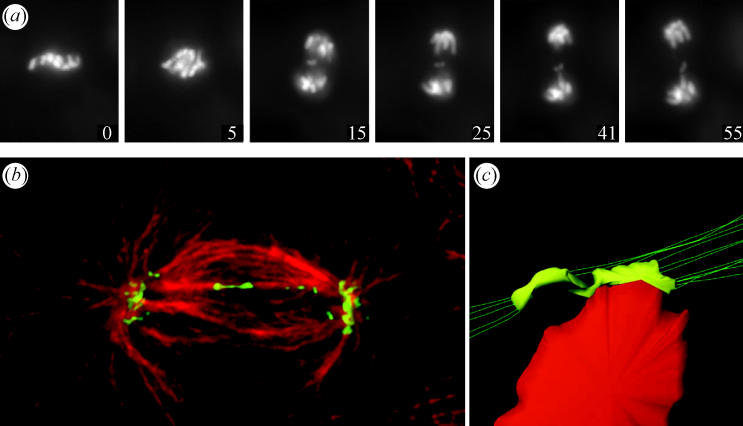
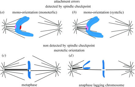
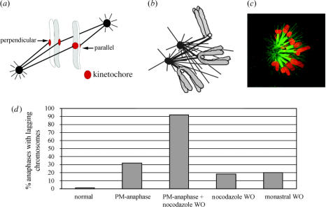
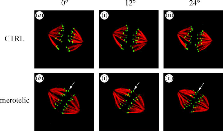
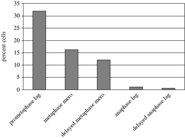
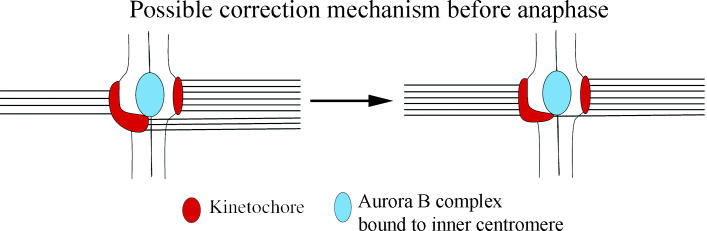
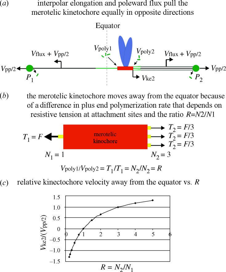
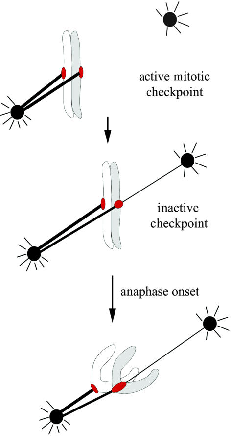
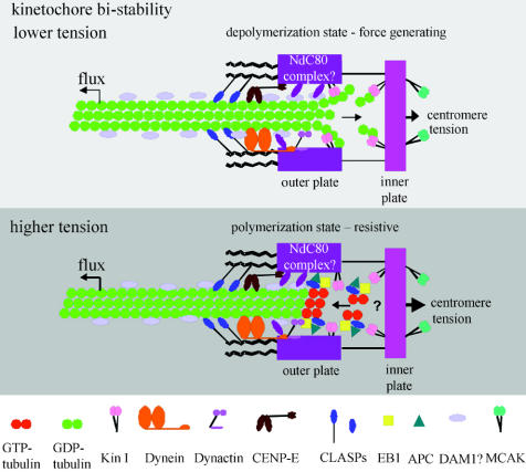
Merotelic kinetochore attachment is a major source of aneuploidy in mammalian tissue cells in culture. Mammalian kinetochores typically have binding sites for about 20-25 kinetochore microtubules. In prometaphase, kinetochores become merotelic if they attach to microtubules from opposite poles rather than to just one pole as normally occurs. Merotelic attachments support chromosome bi-orientation and alignment near the metaphase plate and they are not detected by the mitotic spindle checkpoint. At anaphase onset, sister chromatids separate, but a chromatid with a merotelic kinetochore may not be segregated correctly, and may lag near the spindle equator because of pulling forces toward opposite poles, or move in the direction of the wrong pole. Correction mechanisms are important for preventing segregation errors. There are probably more than 100 times as many PtK1 tissue cells with merotelic kinetochores in early mitosis, and about 16 times as many entering anaphase as the 1% of cells with lagging chromosomes seen in late anaphase. The role of spindle mechanics and potential functions of the Ndc80/Nuf2 protein complex at the kinetochore/microtubule interface is discussed for two correction mechanisms: one that functions before anaphase to reduce the number of kinetochore microtubules to the wrong pole, and one that functions after anaphase onset to move merotelic kinetochores based on the ratio of kinetochore microtubules to the correct versus incorrect pole.


Figures

Figure 1
Figure 1
Merotelic kinetochores can produce lagging chromosomes near the spindle equator in anaphase. (a) Live-cell imaging of a PtK1 cell expressing GFP-H2B. A lagging chromosome remains near the spindle equator as the rest of the chromosomes segregate to their poles. Time on each frame given in minutes. From Cimini et al. (2002). (b) Spinning disk confocal fluorescence microscopy of PtK1 cell showing how microtubule attachments to the opposite poles stretch a merotelic kinetochore laterally from its normal width of about 0.4 to 2 μm or more. The kinetochores (green) were labelled by immunofluorescence with CREST antibodies to inner kinetochore proteins while microtubules (red) were labelled with tubulin antibodies. From Cimini et al. (2001). (c) 3D reconstruction from electron micrographs of a merotelic kinetochore of an anaphase lagging chromosome done by Alexey Khodjakov. From Cimini et al. 2001.
Figure 2
Figure 2
Attachment errors of kinetochores to spindle microtubules. The intensity of red at the kinetochores is an indication of their spindle checkpoint activity. See text for details.
Figure 3
Figure 3
Some factors affecting merotelic kinetochore formation during prometaphase (ac) and affecting the percent cells with lagging chromosomes in anaphase (d). See text for details. (a) Two different sister chromatid orientations relative to the spindle pole-to-pole axis; one favours merotelic attachment, one does not. From Cimini et al. (2003). (b) Centrosomes often come close together in nocodazole-treated mitotic cells and centrosome separation is slow compared with microtubule polymerization after nocodazole washout producing opportunities for merotelic attachments when the centres separate. (c) Spinning disk confocal fluorescence image of a prometaphase PtK1 cell treated with monastrol to prevent centrosome separation before fixation. Note: kinetochore attachments to both poles and syntelic attachments occur in monastrol blocked cells; both errors can lead to merotelic attachments after drug washout and subsequent centre separation as described in the text. Tubulin immunofluorescence is green while chromosome fluorescence is red. From Canman et al. (2003). (d) The frequency of anaphase cells with lagging chromosomes is substantially increased by experimental treatments that induce premature anaphase or prevent centrosome separation before bipolar spindle assembly or both.
Figure 4
Figure 4
Projection views at three different angles through 3D reconstructions of two metaphase PtK1 spindles (a and b) that were fixed and immunofluorescently labelled with CREST antibodies to the kinetochore (green) and tubulin antibodies to stain kinetochore fibre microtubules (red). Cells had progressed normally through mitosis to metaphase before fixation. Note the cell in b has a merotelic kinetochore shifted toward the equator in between the other pairs of sister kinetochores (arrows). From Cimini et al. (2003).
Figure 5
Figure 5
Merotelic kinetochore formation occurs frequently in early prometaphase and merotelic kinetochores are only partially corrected by late metaphase or by prolonging metaphase by 2 h. Only 1 out of 16 merotelic kinetochores entering normal anaphase produce lagging chromosomes, and this reduction is enhanced by prolonging metaphase. See text for details.
Figure 6
Figure 6
Microtubules attached to the wrong pole may bring their kinetochore attachment sites closer to the Aurora B kinase, which is located within the inner centromere region between sister kinetochores before anaphase. From Cimini et al. (2003).
Figure 7
Figure 7
Live imaging of merotelic kinetochores (green) and spindle microtubules (red) in PtK1 cells using a spinning disk confocal fluorescence microscope as described in the text. Frames are from time-lapse movies of different cells. (a) Metaphase aligned sister chromatid pair near the equator with one merotelic kinetochore (arrow). (b) Merotelic kinetochore in anaphase remained near the spindle equator with the brightness of its kinetochore fibres to opposite poles nearly equal and fibre polymerization nearly equal (R∼1). (c) Merotelic kinetochore in anaphase moved away from the equator in the direction of its brighter kinetochore fibre to the correct pole as the dimmer fibre to the opposite pole polymerized much longer (R>1). (d) A merotelic kinetochore in anaphase that became unusually stretched laterally as it moved away from the equator in the direction of the brighter kinetochore fibre (R>1).
Figure 8
Figure 8
Comparison of the changes in length of kinetochore fibres and interpolar distance during anaphase in PtK1 cells for cells with only normally oriented kinetochores and for cells with a merotelic kinetochore in anaphase as described by Cimini et al. (2004).
Figure 9
Figure 9
Model for how anaphase spindle and kinetochore mechanics move chromosomes with merotelic kinetochores in anaphase. See text for details.
Figure 10
Figure 10
Hypothesis for how syntelic attachment of both sister kinetochores to one pole in prometaphase before merotelic kinetochore formation and bi-orientation can lead to both sisters moving to the same pole in anaphase by the mechanism described in figure 9. Chromosome non-disjunction occurs because the merotelic kinetochore in anaphase has kinetochore microtubules to the same pole as its sister kinetochore with a number that is larger than the number to the opposite, correct pole (R<1).
Figure 11
Figure 11
A model for molecular organization at a vertebrate kinetochore microtubule attachment site. Kinetochores exhibit bi-stability, switching to depolymerization at low attachment site tensions and switching to polymerization at high attachment site tensions (Inoué & Salmon 1995; Rieder & Salmon 1998; Mitchison & Salmon 2001; Maiato et al. 2004). These two persistent states are probably dependent on the dynamic instability of microtubule plus ends. At depolymerizing ends, GDP-dimers within the microtubule lattice lose their lateral connections and their protofilaments curve inside-out promoting dimer dissociation. Energy for producing this curvature is provided by the hydrolysis of GTP that occurred previously during polymerization. The depolymerization state of the kinetochore is force generating and pulls the attachment site in a minus direction along the microtubule lattice coupled to depolymerization within the attachment site. An attachment site appears to switch to the polymerization state when attached plus ends of microtubules switch to polymerization. Polymerizing ends are capped and stabilized by unhydrolysed GTP-tubulin dimer, the polymerizing subunit. Polymerizing ends rarely push kinetochores during mitosis (Rieder & Salmon 1998), but attachment sites with polymerizing ends generate resistive tension to pulling forces produced by poleward microtubule flux, spindle interpolar elongation and centromere stretch. The Ndc80 complex of proteins is required to organize kinetochore outer plate attachment site structure and, based on budding yeast studies, probably provides dynamic linkage (projection in drawing) between the outer plate and microtubule associated proteins on the microtubule lattice similar to the DAM1 complex in yeast. Kin I kinesins promote depolymerization at depolymerizing ends and the Kin I kinesin MCAK at the inner centromere may assist in eliminating merotelic kinetochore attachments. Cytoplasmic dynein, dynactin and the kinesin CENP-E are components of the kinetochore corona. Dynein minus-motor activity may enhance depolymerase activity by pulling the microtubule lattice into the attachment site. CENP-E and dynein have major functions in the spindle checkpoint (Cleveland et al. 2003). Polymerizing ends concentrate EB1 at their tips, which stabilizes growth and antagonizes the depolymerases. Adenomatous polyposis coli (APC) protein binds EB1 and may help link polymerizing ends at kinetochores. Another protein that binds polymerizing ends is CLASP1, and CLASP1 is required to prevent persistent depolymerization (Maiato et al. 2004). Not shown in the drawing are potential roles of Cdc42 and mDia3 in linking APC, EB1 and microtubules to kinetochores (Green & Kaplan 2003; Yasuda et al. 2004) and RanBP1 and RanGap for stable kinetochore microtubule attachment (reviewed in Maiato et al. 2004). Drawing is modified from Maiato et al. (2004).

Similar articles

Cited by

References

    1. Alexander S.P, Rieder C.L. Chromosome motion during attachment to the vertebrate spindle: initial saltatory-like behaviour of chromosomes and quantitative analysis of force production by nascent kinetochore fibers. J. Cell Biol. 1991;113:805–815. - PMC - PubMed
    1. Bharadwaj, R., Qi, W. & Yu., H. T. 2004 Identification of two novel components of the human NDC80 kinetochore complex. J. Biol. Chem.279, 13076–13085. - PubMed
    1. Biggins S, Murray A.W. The budding yeast protein kinase Ipl1/Aurora allows the absence of tension to activate the spindle checkpoint. Genes Dev. 2001;15:3118–3129. - PMC - PubMed
    1. Biggins S, Severin F.F, Bhalla N, Sassoon I, Hyman A.A, Murray A.W. The conserved protein kinase Ipl1 regulates microtubule binding to kinetochores in budding yeast. Genes Dev. 1999;13:532–544. - PMC - PubMed
    1. Brinkley, B. R. & Cartwright, Jr., J. 1975 Cold-labile and cold-stable microtubules in the mitotic spindle of mammalian cells. Ann NY Acad. Sci.253, 428–439. - PubMed

Publication types

LinkOut - more resources