. 2005 Mar 29;360(1455):637-47, discussion 447-8.
doi: 10.1098/rstb.2004.1618.

Explaining the oligomerization properties of the spindle assembly checkpoint protein Mad2

Affiliations

Explaining the oligomerization properties of the spindle assembly checkpoint protein Mad2

Anna DeAntoni et al. Philos Trans R Soc Lond B Biol Sci. .

Abstract

Mad2 is an essential component of the spindle assembly checkpoint (SAC), a molecular device designed to coordinate anaphase onset with the completion of chromosome attachment to the spindle. Capture of chromosome by microtubules occur on protein scaffolds known as kinetochores. The SAC proteins are recruited to kinetochores in prometaphase where they generate a signal that halts anaphase until all sister chromatid pairs are bipolarly oriented. Mad2 is a subunit of the mitotic checkpoint complex, which is regarded as the effector of the spindle checkpoint. Its function is the sequestration of Cdc20, a protein required for progression into anaphase. The function of Mad2 in the checkpoint correlates with a dramatic conformational rearrangement of the Mad2 protein. Mad2 adopts a closed conformation (C-Mad2) when bound to Cdc20, and an open conformation (O-Mad2) when unbound to this ligand. Checkpoint activation promotes the conversion of O-Mad2 to Cdc20-bound C-Mad2. We show that this conversion requires a C-Mad2 template and we identify this in Mad1-bound Mad2. In our proposition, Mad1-bound C-Mad2 recruits O-Mad2 to kinetochores, stimulating Cdc20 capture, implying that O-Mad2 and C-Mad2 form dimers. We discuss Mad2 oligomerization and link our discoveries to previous observations related to Mad2 oligomerization.


Figures

Figure 1
Figure 1
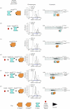
Schematic view of the Mad2 exchange model and the Mad2 template model. (a) The Mad2 exchange model predicts that in the presence of Cdc20 the Mad1/C-Mad2 core complex, a 2 : 2 tetramer (Luo et al. 2002; Sironi et al. 2002) dissociates and releases Mad2 for Cdc20 binding. A Cdc20/C-Mad2 complex is formed and a C-Mad2 vacancy is left on Mad1. This vacancy will be filled by cytosolic O-Mad2. An implication of this model is that Mad1 and Cdc20 compete for Mad2 binding, because both contain a Mad2-binding motif. (b) The Mad1/Mad2 tetramer is stable in the Mad2 template model (De Antoni et al. 2005). C-Mad2 on this tetramer acts as a receptor for cytosolic O-Mad2. Thus, a C-Mad2/O-Mad2 oligomer is formed at kinetochores, and O-Mad2 within this oligomer is a source of Mad2 for Cdc20, which leaves an O-Mad2 vacancy (but not a C-Mad2 vacancy as in the exchange model).
Figure 2
Figure 2
Opposite. The oligomerization state of Mad2 can be explained as an interaction of the two conformers of Mad2, O-Mad2 and C-Mad2. (a) The elution profile of bacterially expressed Mad2wt was typical of a Mad2 oligomer, in agreement with previous reports (Fang et al. 1998; Luo et al. 2000, 2002; Sironi et al. 2001). We interpret this result as the formation of a complex between O-Mad2 and empty C-Mad2 forming in bacterial preparations of Mad2. (b) When challenged with a Cdc20 peptide, Mad2 is turned into a monomeric Cdc20-bound C-Mad2 species, which agrees with previous reports (Fang et al. 1998; Luo et al. 2000, 2002; Sironi et al. 2001). This experiment shows that the accumulation of C-Mad2 reverts the oligomerization of Mad2. To generate Mad2/Cdc20, Mad2wt was incubated for 1 h with a 10 fold excess of a synthetic peptide corresponding to residues between 111 and 138 of Cdc20 (Cdc20111–138). This segment is a stronger Mad2 ligand than full-length Cdc20, probably because the Mad2-binding region is partially buried in full-length Cdc20 (Tang et al. 2001; Zhang & Lees 2001). The resulting Mad2/Cdc20 complex runs as a Mad2 monomer because the contribution of the Cdc20 peptide to the overall shape and molecular weight of Mad2 is near to negligible. (c) Mad2ΔC is locked in the O-Mad2 conformation and is also monomeric, which agrees with previous observations (Fang et al. 1998; Luo et al. 2000, 2002; Sironi et al. 2001). (d) When O-Mad2ΔC and Cdc20-bound C-Mad2wt (generated as described in panel b) are mixed in roughly equimolar amounts, a stoichiometric complex form that runs very similarly to the Mad2wt oligomers. (e) The existence of C-Mad2 in Mad2wt is supported by partial binding of Mad2ΔC to Mad2wt also in the absence of Cdc20 peptide. The shift in elution volume of Mad2ΔC caused by its incorporation into a complex with C-Mad2wt can be appreciated by a comparison with panel c. Some monomeric Mad2wt, presumably in the O-Mad2 conformation, is competed by Mad2ΔC off the wild-type oligomer and it appears as a monomeric species. The Mad2 proteins used in panels ae were purified and analysed by SEC on a Superdex-75 PC 3.2/30 column essentially as described (Sironi et al. 2001, 2002; De Antoni et al. 2005). For every chromatogram, pure proteins were loaded onto the column and the contents of 12 consecutive 30 μl fractions eluting between 0.96 and 1.32 ml were analysed by SDS-PAGE and stained with Coomassie.
Figure 3
Figure 3
Oligomeric state of different forms of Mad2 after purification and in cell extracts. The elution of bacterially expressed and purified monomeric and oligomeric Mad2 proteins was used to discern the oligomerization state of Mad2 in HeLa cell extracts. Protein samples were loaded on a Superose-12 PC 3.2/30 column equilibrated in 50 mM Hepes pH 7.5, 150 mM NaCl, 1 mM DTT, 5 mM EDTA. Elution was carried out at 40 μl min−1. For each run, identical 35 μl fractions were collected and then subjected to SDS-PAGE, followed by Coomassie staining or western blotting. Arrows indicated the position of the peaks of three molecular weight markers. (a) SDS-PAGE analysis of monomeric O-Mad2ΔC. (b) SDS-PAGE analysis of monomeric O-Mad2wt. (c) HeLa cells were harvested by scraper detaching and lysed in 50 mM Hepes pH 7.5, 150 mM NaCl, 1 mM DTT, 5 mM EDTA, 50 mM NaF, 20 mM Na4-pyrophosphate, 0.5 μM okadaic acid, 100 ng ml−1 leupeptine, 100 ng ml−1 aprotinin, 1 mM PMSF, protease inhibitors (Roche), 0.1% NP40 for 20 min on ice. Cell lysates were clarified by centrifugation for 15 min at 13 000 r.p.m. at 4 °C in an Eppendorf microcentrifuge. Cell lysates (70 μg) were subjected to SEC analysis.
Figure 4
Figure 4
Opposite. Behaviour of the Mad2R133A mutant. (a) Mad2R133A is monomeric because the R133A mutation (yellow triangle) impairs the interaction between O- and C-Mad2. (b) Mad2R133A/Cdc20 runs as a 1:1 Mad2/peptide complex. (c) Mad2ΔC-R133A is monomeric and locked as O-Mad2, like Mad2ΔC, owing to the ΔC deletion. (d) O-Mad2ΔC-R133A does not bind C-Mad2R133A/Cdc20 and the individual profiles of the monomers largely overlap, although a small shift is observed, possibly revealing small residual binding. (e) When the R133A mutation is present only on the C-Mad2 moiety, an interaction with O-Mad2 is observed. Note that the elution of this complex is shifted to the lower molecular weights relative to Mad2wt (see figure 2a,d). (f) As in f, the R133A mutation is present on only one face of the interaction, which, in this case, is the O-Mad2 moiety. As in panel e, an interaction with C-Mad2 is observed. The Mad2 proteins used in panels af were purified and analysed by SEC on a Superdex-75 PC 3.2/30 column essentially as described (Sironi et al. 2001, 2002; De Antoni et al. 2005). For every chromatogram, pure proteins were loaded onto the column and the contents of 12 consecutive 30 μl fractions eluting between 0.96 and 1.32 ml were analysed by SDS-PAGE and stained with Coomassie.
Figure 5
Figure 5
Summary of Mad2 oligomerization mechanism. Although neither conformer interacts with itself, the idea that the oligomerization of Mad2 is caused by an interaction of O-Mad2 with C-Mad2 explains all observations regarding the oligomerization state of Mad2. In particular, pure preparations of C-Mad2 (panel a) are monomeric. We show this in figure 2b, but this effect of Cdc20 and Mad1 binding on Mad2 has also been described before (Luo et al. 2000, 2002, 2004; Sironi et al. 2001, 2002; De Antoni et al. 2005). Similarly, pure O-Mad2ΔC is monomeric (panel b), a result that has also already been discussed (Fang et al. 1998; Luo et al. 2000, 2002, 2004; Sironi et al. 2001, 2002; De Antoni et al. 2005). In panels c and d, we summarize our finding that the oligomerization of Mad2 can be explained as an interaction of O-Mad2 with C-Mad2. The difference between panels c and d is the presence of a Mad2 ligand, which will cure the polydispersity of oligomeric preparations of empty Mad2 (Sironi et al. 2001; De Antoni et al. 2005; Luo et al. 2004). Recently, we have shown that the interaction of O-Mad2 with C-Mad2, which we have ‘simulated’ here with purified components, is essential for Mad2 recruitment to the kinetochore and maintenance of the spindle checkpoint (De Antoni et al. 2005). Finally, we postulate that the reason why the R133A mutant is monomeric is that this residue lies at the interface between the O-Mad2 and C-Mad2 monomers (panel e).

Similar articles

Cited by

References

    1. Aravind L, Koonin E.V. The HORMA domain: a common structural denominator in mitotic checkpoints, chromosome synapsis and DNA repair. Trends Biochem. Sci. 1998;23:284–286. - PubMed
    1. Bharadwaj R, Yu H. The spindle checkpoint, aneuploidy, and cancer. Oncogene. 2004;23:2016–2027. - PubMed
    1. Chen R.-H. BubR1 is essential for kinetochore localization of other spindle checkpoint proteins and its phosphorylation requires Mad1. J. Cell Biol. 2002;158:487–96. - PMC - PubMed
    1. Chen R.H, Shevchenko A, Mann M, Murray A.W. Spindle checkpoint protein Xmad1 recruits Xmad2 to unattached kinetochores. J. Cell Biol. 1998;143:283–295. - PMC - PubMed
    1. Chen R.H, Brady D.M, Smith D, Murray A.W, Hardwick K.G. The spindle checkpoint of budding yeast depends on a tight complex between the Mad1 and Mad2 proteins. Mol. Biol. Cell. 1999;10:2607–2618. - PMC - PubMed

Publication types

LinkOut - more resources