. 2005 May 23;169(4):635-46.
doi: 10.1083/jcb.200410081. Epub 2005 May 16.

Essential function of Drosophila Sec6 in apical exocytosis of epithelial photoreceptor cells

Affiliations

Essential function of Drosophila Sec6 in apical exocytosis of epithelial photoreceptor cells

Slobodan Beronja et al. J Cell Biol. .

Abstract

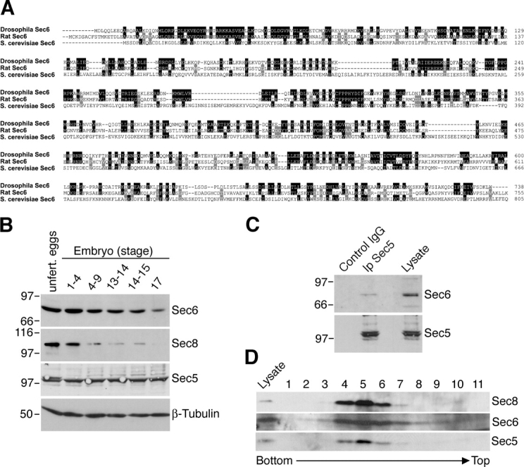
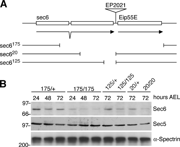
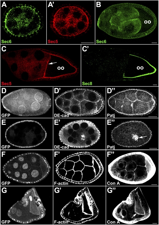
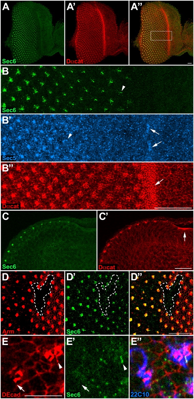
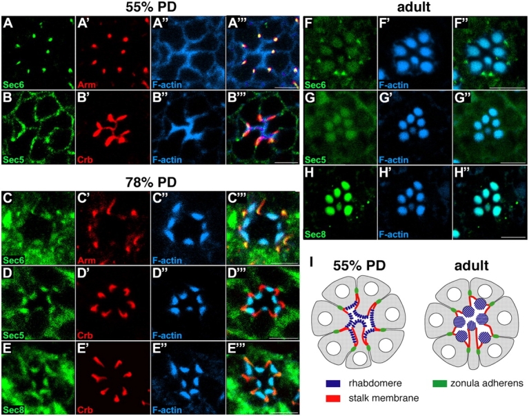
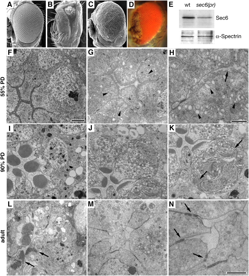
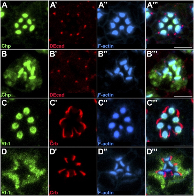
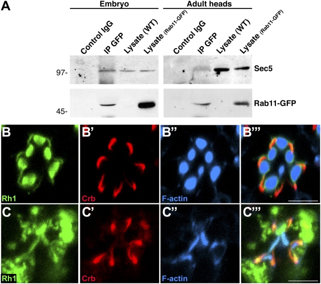
Polarized exocytosis plays a major role in development and cell differentiation but the mechanisms that target exocytosis to specific membrane domains in animal cells are still poorly understood. We characterized Drosophila Sec6, a component of the exocyst complex that is believed to tether secretory vesicles to specific plasma membrane sites. sec6 mutations cause cell lethality and disrupt plasma membrane growth. In developing photoreceptor cells (PRCs), Sec6 but not Sec5 or Sec8 shows accumulation at adherens junctions. In late PRCs, Sec6, Sec5, and Sec8 colocalize at the rhabdomere, the light sensing subdomain of the apical membrane. PRCs with reduced Sec6 function accumulate secretory vesicles and fail to transport proteins to the rhabdomere, but show normal localization of proteins to the apical stalk membrane and the basolateral membrane. Furthermore, we show that Rab11 forms a complex with Sec5 and that Sec5 interacts with Sec6 suggesting that the exocyst is a Rab11 effector that facilitates protein transport to the apical rhabdomere in Drosophila PRCs.


Figures

Figure 1.
Figure 1.
Drosophila Sec6 is ubiquitously expressed and interacts with Sec5. (A) Protein sequence alignment of Drosophila, rat, and yeast Sec6. (B) Developmental immunoblot detecting Sec6, Sec8, and Sec5 in unfertilized eggs and embryos. Sec6 (81 kD) is present in unfertilized eggs and at decreasing levels throughout embryogenesis. The amount of Sec5 remains constant. Sec8 (107 kD) is also maternally provided, but its expression strongly decreases and is undetectable by the end of embryogenesis. (C) Co-IP using Sec5 mAb 16A2 precipitates Sec6 as detected by immunoblotting. (D) Density gradient cosedimentation of membranes shows that Sec6, Sec5, and Sec8 largely cofractionate. Note that Sec6 is found in more fractions than either Sec5 or Sec8.
Figure 2.
Figure 2.
Generation of sec6 mutations. (A) Extent of deletions sec6 175, sec6 20, and sec6 125 induced by imprecise excision of the EP2021 P element that maps between sec6 and Eip55E. (B) Zygotic mutants for all three sec6 alleles show a decrease of Sec6 whereas levels of Sec5 are not affected. AEL, after egg laying.
Figure 3.
Figure 3.
Sec6 is required for plasma membrane integrity of the female germline. (A) Sec6 and Sec5 show a punctate cytoplasmic distribution in the germline of early follicles. (B) Stage 9 follicle showing uniform, punctate distribution of Sec6 in nurse cells and the follicular epithelium and lower levels in the oocyte (oo). (C) Stage 9 follicle showing Sec8 and Sec5 accumulation at the oocyte membrane. Note that Sec8 levels in nurse cells and follicle cells are very low compared with Sec5 or Sec6 (B), and that Sec8 is not detected at the anterior oocyte membrane in contrast to Sec5 (arrow). (D and F) Wild-type follicle. (E and G) Follicles with a sec6 20 mutant germline clone (E) or partially mutant germline clone (G) as identified by the absence of GFP. DEcad, Patj, F-actin, and Con A do not accumulate between Sec6 mutant germline cells suggesting that plasma membranes are absent. Note that in the partial germline clone in G, plasma membrane is lost except around the oocyte (arrow) and a remnant of membrane between the two GFP-positive nurse cell nuclei (arrowhead). A central clump of material highlighted by DEcad (E′) and Patj (E′′) presumably represents collapsed ring canals (arrow). Bars, 20 μm.
Figure 4.
Figure 4.
Distribution of Sec6 in third larval instar eye discs requires Arm. (A) Sec6 and Dαcat colocalize in the third larval instar eye disc. (B) Top view and (C) side view of a portion of a third larval instar eye imaginal disc (A′′, box) showing ∼20 rows of PRC clusters posterior to the morphogenetic furrow (MF; B′′ and C′, arrows). The MF and PRCs show strong apical enrichment of Dαcat at the ZA. Sec6 colocalizes with Dαcat at the ZA two to four rows behind the MF (B, arrowhead). Some apical accumulation of Sec5 is already apparent in the MF (B′, arrows). However, within the PRC clusters, apical Sec5 accumulation is trailing that of Sec6 by several rows (B′, arrowhead). Apical Sec5 appears more diffuse than Sec6. (D) Clone of arm XP33 mutant eye disc cells marked by the loss of Arm (outline) does not show ZA enrichment of Sec6 (D′). (E) Clone of arm XP33 mutant eye disc cells marked by the loss of DEcad. PRCs are labeled with a neuronal marker 22C10 (E′′). DEcad (E) and Sec6 (E′) accumulation between wild-type PRCs (E and E′, arrowheads) is not seen at the interface between a wild-type and an arm XP33 mutant cell (E and E′, arrow). All images represent a projection of a 20-μm-deep series of Z-sections. Bars: (A–D) 20 μm; (E) 10 μm.
Figure 5.
Figure 5.
Sec6, Sec5, and Sec8 distribution in pupal and adult PRCs. All panels show optical cross sections through individual wild-type ommatidia at 55% PD (A and B), 78% PD (C–E), and adult (F–H). (A) Sec6 localizes to the ZA (marked by Arm) of PRCs at 55% PD. (B) Sec5 associates with the basolateral and apical membrane at 55% PD, and appears somewhat enriched at the apical membrane were it colocalizes with Crb at the stalk membrane. (C–H) Sec6, Sec5, and Sec8 localize to the rhabdomere, marked by strong labeling of F-actin with phalloidin. Sec6 also remains associated with the ZA, marked by Arm in C. None of the exocyst proteins show significant accumulation at the stalk membrane, marked by Crb in D and E. All three exocyst proteins are also found throughout the PRC cytoplasm. (I) Diagram of cross sections of an ommatidium at 55% PD and adult. The rhabdomere and the stalk membrane comprise the apical membrane of PRCs, whereas the ZA and the rest of the membrane comprise the basolateral membrane. Bars, 5 μm.
Figure 6.
Figure 6.
Accumulation of secretory vesicles in sec6(pr) mutant PRCs. (A–C) Scanning EM of a wild-type (A), sec6 20 (B), and sec6(pr) mutant eyes (C). (D) Adult sec6(pr) mutant eye showing loss of pigment in the anterior. (E) Immunoblot showing that sec6(pr) mutant adult retinas have strongly decreased levels of Sec6 compared with wild type. (F–N) Transmission EM of wild-type (F and I) and sec6(pr) mutant PRCs (G, H, J–N) at 55% PD (F–H), 90% PD (I–K), and adult (L–N). sec6(pr) mutant PRCs show prominent groups of small vesicles (arrowheads) in the apical cytoplasm at 55% PD and gaps in the array of microvilli are seen (G and H; arrow). In wild type, such vesicles are rare and microvilli form a continuous rim (F). sec6(pr) mutant PRCs at 90% PD are filled with vesicles 100–300 μm in diameter and PRCs appear swollen (J and K) in contrast to wild type (I). Rhabdomeres in sec6(pr) mutant PRCs are small and flattened and some cells display an enlarged ER (K, arrows). Adult sec6(pr) mutant PRCs show strong reduction in size (L, arrows) or complete loss of rhabdomeres (M), but display normal ZAs (N, arrows). Bars: (F, G, and I–M) 1 μm; (H and N) 0.5 μm.
Figure 7.
Figure 7.
Sec6 is required for protein transport to the rhabdomere. Optical cross sections of individual wild-type (A and C) and sec6(pr) ommatidia (B and D) at 78% PD (A and B) or from 1-d-old adult eye (C and D). Chp (B) and Rh1 (D) accumulate in the cytoplasm of sec6(pr) mutant PRCs in contrast to wild type, whereas DEcad and Crb show normal subcellular distributions. Rhabdomeres are identified by labeling F-actin with phalloidin. Bars, 5 μm.
Figure 8.
Figure 8.
The exocyst is a Rab11 effector and mediates direct Rh1 transport to the rhabdomere. (A) Co-IP with anti-GFP mAb precipitates Sec5 from lysates of da-Gal4 UAS-Rab11-GFP embryos ubiquitously expressing Rab11::GFP and Rh1-Gal4 UAS-Rab11-GFP eyes expressing Rab11::GFP in PRCs R1-R6. (B) PRCs of shi ts2 adults grown at 29°C for 3 d show normal accumulation of Rh1 in the rhabdomeres. (C) PRCs of shi ts2 sec6(pr) flies grown at 29°C for 3 d show cytoplasmic accumulation of Rh1. Bars, 5 μm.

Similar articles

Cited by

References

    1. Andrews, H.K., Y.Q. Zhang, N. Trotta, and K. Broadie. 2002. Drosophila sec10 is required for hormone secretion but not general exocytosis or neurotransmission. Traffic. 3:906–921. - PubMed
    1. Ang, A.L., T. Taguchi, S. Francis, H. Folsch, L.J. Murrells, M. Pypaert, G. Warren, and I. Mellman. 2004. Recycling endosomes can serve as intermediates during transport from the Golgi to the plasma membrane of MDCK cells. J. Cell Biol. 167:531–543. - PMC - PubMed
    1. Ayscough, K.R., J. Stryker, N. Pokala, M. Sanders, P. Crews, and D.G. Drubin. 1997. High rates of actin filament turnover in budding yeast and roles for actin in establishment and maintenance of cell polarity revealed using the actin inhibitor latrunculin-A. J. Cell Biol. 137:399–416. - PMC - PubMed
    1. Bucci, C., R.G. Parton, I.H. Mather, H. Stunnenberg, K. Simons, B. Hoflack, and M. Zerial. 1992. The small GTPase rab5 functions as a regulatory factor in the early endocytic pathway. Cell. 70:715–728. - PubMed
    1. Chu, D.T., and M.W. Klymkowsky. 1989. The appearance of acetylated alpha-tubulin during early development and cellular differentiation in Xenopus. Dev. Biol. 136:104–117. - PubMed

Publication types

MeSH terms