. 2005 May 23;169(4):569-76.
doi: 10.1083/jcb.200501071. Epub 2005 May 16.

Inducible ablation of mouse Langerhans cells diminishes but fails to abrogate contact hypersensitivity

Affiliations

Inducible ablation of mouse Langerhans cells diminishes but fails to abrogate contact hypersensitivity

Clare L Bennett et al. J Cell Biol. .

Abstract

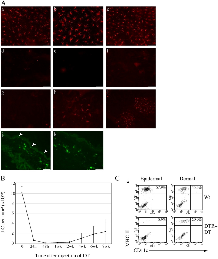
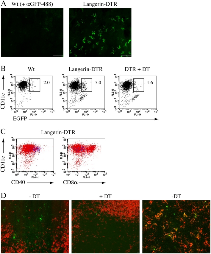
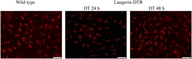
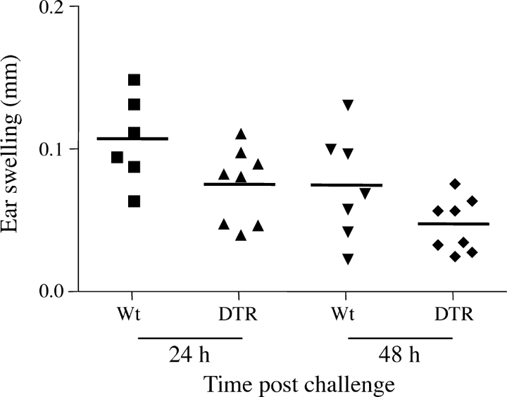
Langerhans cells (LC) form a unique subset of dendritic cells (DC) in the epidermis but so far their in vivo functions in skin immunity and tolerance could not be determined, in particular in relation to dermal DC (dDC). Here, we exploit a novel diphtheria toxin (DT) receptor (DTR)/DT-based system to achieve inducible ablation of LC without affecting the skin environment. Within 24 h after intra-peritoneal injection of DT into Langerin-DTR mice LC are completely depleted from the epidermis and only begin to return 4 wk later. LC deletion occurs by apoptosis in the absence of inflammation and, in particular, the dDC compartment is not affected. In LC-depleted mice contact hypersensitivity (CHS) responses are significantly decreased, although ear swelling still occurs indicating that dDC can mediate CHS when necessary. Our results establish Langerin-DTR mice as a unique tool to study LC function in the steady state and to explore their relative importance compared with dDC in orchestrating skin immunity and tolerance.


Figures

Figure 1.
Figure 1.
Generation of Langerin-DTR mice. (A) Schematic representation of the targeting strategy used for the generation of Langerin-DTR mice. HR, homologous recombination; K, KpnI; E, EcoRI; N, NotI. (B) Identification of targeted ES cell clones by Southern hybridization using probe [1] after digestion with KpnI, 8-kb wt allele and 3-kb targeted allele. +/+, wt; +/t, targeted heterozygote. (C) Depletion of the NeoR cassette was verified by Southern blot analysis using the DTR:GFP cassette as probe [2]: +, Langerin-DTR founder; wt, wt control; ΔNeo, Langerin-DTRΔNeo. (D) Genomic PCR of Langerin-DTR mice, lanes 1–5, Langerin-DTR mutant and wt littermates; +, Langerin-DTR founder; –, water control.
Figure 2.
Figure 2.
Efficient and specific ablation of LC from Langerin-DTR mice. (A) Epidermal sheets were separated from mouse ears and stained for MHCII to visualize LC: (a) wt; (b) wt + DT (72 h); (c) Langerin-DTR. Mutant mice were subsequently injected i.p. with DT and LC visualized to determine the kinetics of depletion and repopulation; (d) 24 h; (e) 48 h; (f) 2 wk; (g–i) 4 wk. Pictures are representative of four different mice for each time point, γ adjustments were made to reduce background on epidermal sheets. Cross sections through ear skin were stained with MHCII to demonstrate selective depletion of LC and an intact dDC compartment after DT. (j) Langerin-DTR; (k) DTR + DT (24 h). Arrowheads identify MHCII+ LC in the epidermis. Bars, 50 μm. (B) Quantification of depletion and repopulation of LC after injection of DT. Numbers of MHCII+ LC were counted using a gridded eyepiece with the 40× objective. 20 fields of evenly spaced cells were counted from four different mice for each time point. At 6 wk after injection of DT, LC numbers are still significantly decreased compared with wt mice (Mann-Whitney test wt vs. Langerin-DTR mice at 6 wk: P = 0.0286). Error bars represent SD. (C) LC but not dDC are ablated in Langerin-DTR mice. Emigrant cells from the epidermis or dermis of wt and Langerin-DTR mice injected with DT 48 h earlier were analyzed by flow cytometry. Numbers show the percentage of CD11c/MHCII double-positive cells, FACS plots are representative of three separate experiments.
Figure 3.
Figure 3.
EGFP expression in Langerin-DTR mice. (A) Expression of EGFP in Langerin-DTR epidermal sheets. Visualization of EGFP expression was enhanced by incubation of an anti–GFP-488 antibody, which specifically labeled cells from Langerin-DTR mice. Bars, 50 μm. (B) EGFP expression in skin draining LN. CD11c+ LN cells from wt, Langerin-DTR, and Langerin-DTR mice injected with DT (48 h) were enriched and analyzed for EGFP expression, 3.34% ± 0.375 (mean ± SEM, n = 4) of LN cells were gated EGFP-positive compared with 1.47% ± 0.121 (n = 4) background fluorescence in wt mice. Mann-Whitney test wt vs. Langerin-DTR, P = 0.0159. FACS plots are representative of four different experiments. (C) Cells from Langerin-DTR mice were analyzed for expression of surface markers and CD11c. Blue dots are gated EGFP+ cells from B. Plots are representative of at least five different experiments. (D) LNs from Langerin-DTR mice or Langerin-DTR mice injected with DT for 48 h were sectioned and analyzed for the presence of EGFP+ LC. All sections were stained with anti–GFP-488 in addition to anti-B220 (a and b) or anti-MHCII (c) to demarcate B and T cell areas, respectively. Bars, 10 μm.
Figure 4.
Figure 4.
Depletion of LC in the absence of inflammation. Epidermal sheets from wt and Langerin-DTR mice injected with DT were stained with an antibody against the γδ TCR to visualize dendritic epidermal γδ T cells. Pictures are representative of four different mice for each time point. γ adjustments were made to reduce background on epidermal sheets. Bars, 50 μm.
Figure 5.
Figure 5.
Depletion of LC results in sub-optimal CHS responses. Wt or Langerin-DTR mice (n = 6–8) injected with DT at day −3, were sensitized on the abdomen with 1% TNCB and challenged 5 d later with 0.5% TNCB on one ear. Swelling was measured as the difference between the challenged and nonchallenged ear. Ear swelling responses were compared using a repeated measures ANOVA; wt vs. DTR + DT: P = 0.039. One representative experiment out of three is shown. The two additional experiments were performed with four mice per group.

Similar articles

Cited by

References

    1. Allan, R.S., C.M. Smith, G.T. Belz, A.L. van Lint, L.M. Wakim, W.R. Heath, and F.R. Carbone. 2003. Epidermal viral immunity induced by CD8alpha+ dendritic cells but not by Langerhans cells. Science. 301:1925–1928. - PubMed
    1. Anjuere, F., P. Martin, I. Ferrero, M.L. Fraga, G.M. del Hoyo, N. Wright, and C. Ardavin. 1999. Definition of dendritic cell subpopulations present in the spleen, Peyer's patches, lymph nodes, and skin of the mouse. Blood. 93:590–598. - PubMed
    1. Banchereau, J., and R.M. Steinman. 1998. Dendritic cells and the control of immunity. Nature. 392:245–252. - PubMed
    1. Cumberbatch, M., R.J. Dearman, C.E. Griffiths, and I. Kimber. 2003. Epidermal Langerhans cell migration and sensitisation to chemical allergens. APMIS. 111:797–804. - PubMed
    1. Enk, A.H., and S.I. Katz. 1992. Early molecular events in the induction phase of contact sensitivity. Proc. Natl. Acad. Sci. USA. 89:1398–1402. - PMC - PubMed

Publication types

MeSH terms