. 2005 May 16;201(10):1627-35.
doi: 10.1084/jem.20050125.

Fibrinogen and fibronectin binding cooperate for valve infection and invasion in Staphylococcus aureus experimental endocarditis

Affiliations

Fibrinogen and fibronectin binding cooperate for valve infection and invasion in Staphylococcus aureus experimental endocarditis

Yok-Ai Que et al. J Exp Med. .

Abstract

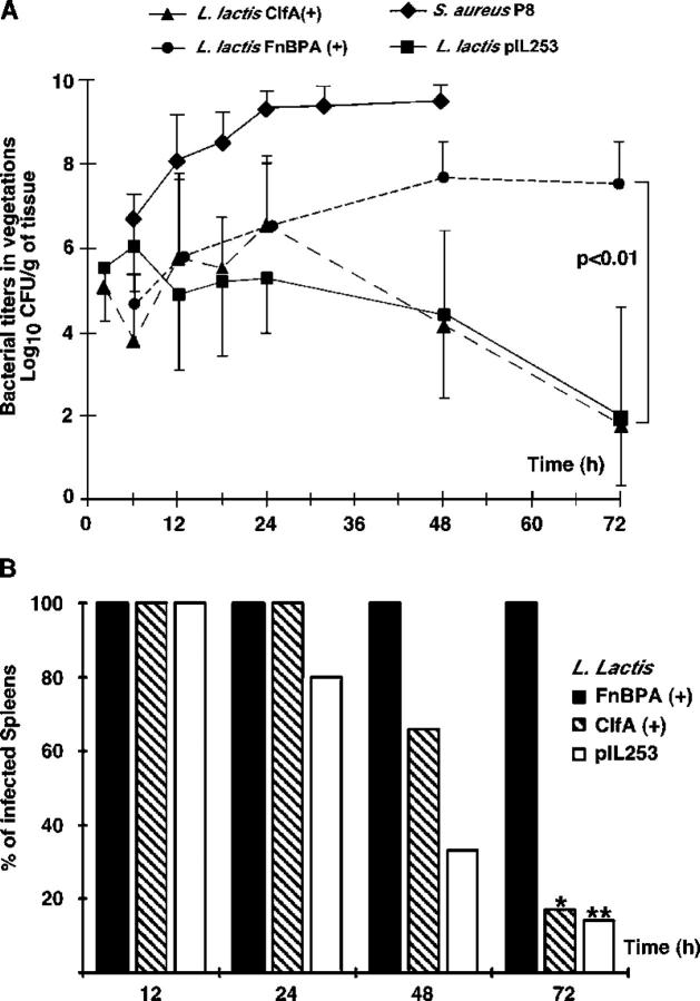
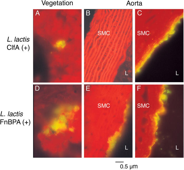
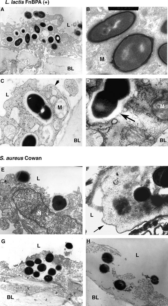
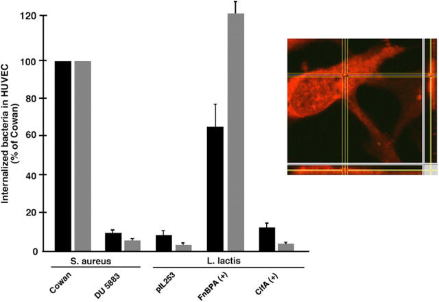
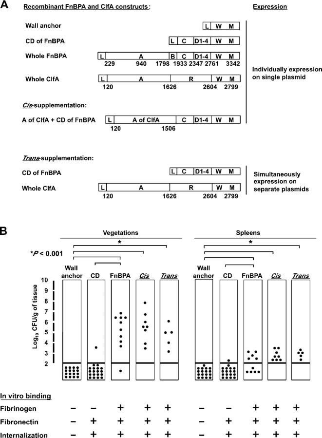
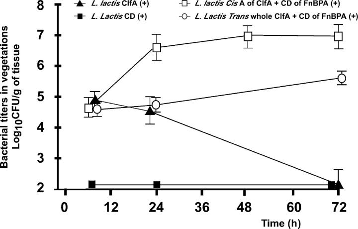
The expression of Staphylococcus aureus adhesins in Lactococcus lactis identified clumping factor A (ClfA) and fibronectin-binding protein A (FnBPA) as critical for valve colonization in rats with experimental endocarditis. This study further analyzed their role in disease evolution. Infected animals were followed for 3 d. ClfA-positive lactococci successfully colonized damaged valves, but were spontaneously eradicated over 48 h. In contrast, FnBPA-positive lactococci progressively increased bacterial titers in vegetations and spleens. At imaging, ClfA-positive lactococci were restricted to the vegetations, whereas FnBPA-positive lactococci also invaded the adjacent endothelium. This reflected the capacity of FnBPA to trigger cell internalization in vitro. Because FnBPA carries both fibrinogen- and fibronectin-binding domains, we tested the role of these functionalities by deleting the fibrinogen-binding domain of FnBPA and supplementing it with the fibrinogen-binding domain of ClfA in cis or in trans. Deletion of the fibrinogen-binding domain of FnBPA did not alter fibronectin binding and cell internalization in vitro. However, it totally abrogated valve infectivity in vivo. This ability was restored in cis by inserting the fibrinogen-binding domain of ClfA into truncated FnBPA, and in trans by coexpressing full-length ClfA and truncated FnBPA on two separate plasmids. Thus, fibrinogen and fibronectin binding could cooperate for S. aureus valve colonization and endothelial invasion in vivo.


Figures

Figure 1.
Figure 1.
Evolution of valve infection in rats with experimental endocarditis caused by various test organisms. Bacterial densities in the vegetations (A) and spleens (B) are indicated. Groups of rats with catheter-induced aortic vegetations were challenged with 10 times the ID80 of the test bacteria and killed at various time points over a period of 3 d. Each data point represents the mean ± SD of vegetation bacterial titers of 8–10 animals. Bacterial strains are indicated on the figure and described in Table I. L. lactis pIL253 carried an empty expression vector (reference 18). L. lactis ClfA (+) and L. lactis FnBPA (+) carried a vector expressing ClfA and FnBPA, respectively (references 18, 47). S. aureus P8 was used as a positive control. Rats infected with L. lactis FnBPA (+) worsened both vegetation bacterial titers and spleen infection (median [range] bacteria/gram of spleen = 3.04 [1.92–3.3], 1.8 [1.29–3.21], 1.92 [1.41-3.2], and 1.84 [1.08–3.21] at 12, 24, 48, and 72 h, respectively) over time. In contrast, rats infected with either L. lactis pIL253 or L. lactis ClfA (+) underwent spontaneous clearing of the infection (* and **, P < 0.01).
Figure 2.
Figure 2.
Immunohistochemistry of the vegetations and neighboring valve endothelium in rats infected with either ClfA-positive (L. lactis) or FnBPA-positive (L. lactis) recombinant lactococci. Animals were killed 24 h after inoculation, and tissues were processed as described previously. Expression of the recombinant adhesins was detected with either anti–ClfA F(ab)'2 fragments (A and B) or anti–FnBPA F(ab)'2 fragments (D and E). C and F were labeled with anti–von Willebrand primary antibodies to determine the presence of the endothelial layer. Fluorescence indicates the presence of bacteria (A, D, and E) and endothelial cells (C and F). Comparison between Gram's stains and immunodetected bacteria indicated that all visible microorganisms expressed their recombinant gene (unpublished data; SMC, smooth muscle cells; L, vascular lumen).
Figure 3.
Figure 3.
Electron microscopy imaging of the valve endothelium adjacent to infected vegetations. Rats were infected with either FnBPA-positive recombinant lactococci (top) or S. aureus Cowan I (bottom). (A) Presentation of an osmium staining of an endothelial cell sandwiched by the vascular lumen (L) on top and the basal lamina (BL) on bottom. Lactococci adherent to the endothelial cell are surrounded by a dark halo, whereas lactococci inside the cell are surrounded by a clear halo. (B) A PAS staining of the same cell. The fact that the clear halo surrounding the intra-endothelial lactococci takes PAS indicates that it consists of extracellular polysaccharides. A mitochondria (M) is observed. (C) An osmium staining of an intracellular dividing lactococcus. The endothelial plasma membrane (arrow) and mitochondria (M) are indicated. (D) Tannic acid staining that helps distinguish between the presence and the absence of intracellular membranes. The membrane lining mitochondria and intracellular vesicles is clearly apparent, whereas no clear membrane surrounds the internalized lactococcus (arrows). E–G depict S. aureus Cowan I at various infection stages: (E) adherence and invasion; (F and G) intracellular location; and (H) cell disruption. E, G, and H are stained by osmium staining and F is stained by tannic acid (arrows, plasma membranes; N, nucleus; L, vascular lumen; BL, basal lamina). Magnification was 8,000–16,000.
Figure 4.
Figure 4.
Internalization of staphylococci and lactococci producing or not producing fibronectin-binding proteins by cultured HUVECs. Internalization was measured both by a penicillin–lysostaphin protection assay (black bars) and by flow cytometry (gray bars; reference 8). The results present the mean ± SD of three independent experiments and are expressed as a percentage of S. aureus Cowan I internalization. Bacterial strains are indicated at the bottom (Table I). The right panel is a magnified view of an internalized lactococcus as detected by confocal microscopy.
Figure 5.
Figure 5.
FnBPA and ClfA constructs expressed in lactococci and used to test the function of specific domains in vitro and in vivo. A presents the nucleotide map of the constructs and B presents the resulting phenotypes. The Wall anchor construct contained only the leader sequence (L) and the wall (W), and membrane (M)-anchoring domains of whole FnBPA. The CD construct contained the leader sequence and the wall-anchoring domains bracketing the C and D regions involved in fibronectin binding. FnBPA, the whole protein containing the A and B additional domains (reference 18); ClfA, the whole S. aureus clumping factor A cloned and expressed in lactococci (R, repeats; reference 47). Construct A-ClfA-CD-FnBPA is a chimera used for cis-supplementation of the construct of the truncated FnBPA (construct CD) with the fibrinogen-binding domain of ClfA. Trans-supplementation was achieved by coexpressing truncated FnBPA (construct CD) with whole ClfA on separate plasmids. The top panel of B presents the vegetation and spleen bacterial densities in rats challenged intravenously with a bacterial number inoculum equivalent to the ID80 of recombinant lactococci expressing whole FnBPA (i.e., 105 CFU) and killed after 24 h (Fig. 1). Each dot represents one individual animal. Mean bacterial densities were notably higher (P < 0.01) in rats infected either with lactococci expressing whole FnBPA or with lactococci expressing the cis- or the trans-supplemented constructs. The bottom panel of B depicts the in vitro adherence and internalization phenotype of the various constructs. ClfA-expressing lactococci were used as a control. −, optical densities (for binding) or bacterial counts (for internalization) less than or equal to the background; +, values greater than or equal to the background.
Figure 6.
Figure 6.
Evolution of valve infection in rats challenged with lactococci expressing truncated FnBPA supplemented or not supplemented with the fibrinogen-binding domain of ClfA in cis or in trans. Recombinants expressing either whole ClfA or truncated FnBPA (construct CD+) are depicted as controls. Each data point represents the mean ± SD (n ≥ 5 individual rats). The slower growth rate of the trans versus cis construct in rats mirrored its slower growth rate in vitro, because of the simultaneous presence of two separate plasmids. Other details are the same as in Fig. 1.

References

    1. Lowy, F.D. 1998. Staphylococcus aureus infections. N. Engl. J. Med. 339:520–532. - PubMed
    1. Foster, T.J., and M. Hook. 1998. Surface protein adhesins of Staphylococcus aureus. Trends Microbiol. 6:484–488. - PubMed
    1. Dinges, M.M., P.M. Orwin, and P.M. Schlievert. 2000. Exotoxins of Staphylococcus aureus. Clin. Microbiol. Rev. 13:16–34. - PMC - PubMed
    1. Arvidson, S. 2000. Extracellular enzymes. Gram-positive Pathogens. V.A. Fischetti, R.P. Novick, J.J. Ferretti, D.A. Portnoy, and J.I. Rood, editors. American Society for Microbiology, Washington, DC. 379–385.
    1. Kintarak, S., S.A. Whawell, P.M. Speight, S. Packer, and S.P. Nair. 2004. Internalization of Staphylococcus aureus by human keratinocytes. Infect. Immun. 72:5668–5675. - PMC - PubMed

Publication types

MeSH terms